. 2022 Jun;25(2):167-175.
doi: 10.1007/s40477-021-00589-5. Epub 2021 Jun 12.

Inflammatory disease of the costotransverse joints: US evaluation in 15 symptomatic patients

Affiliations

Inflammatory disease of the costotransverse joints: US evaluation in 15 symptomatic patients

A Del Chiaro et al. J Ultrasound. 2022 Jun.

Abstract

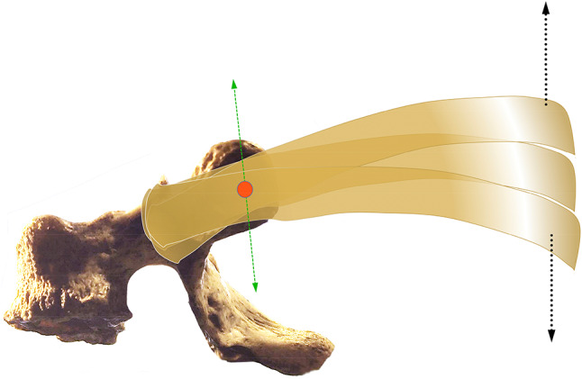
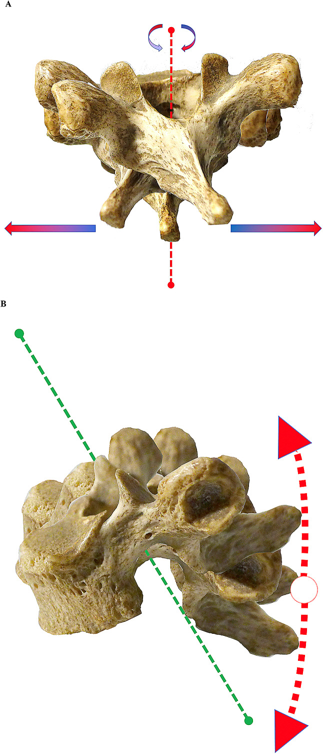
The costotransverse joints (CTJs) are small arthrodial joints which articulate with the costal tuberosity on the transverse process of the thoracic vertebrae. CTJs are composed of oval-shaped facets with a major axis, vertical at the upper vertebrae and almost horizontal at the lower vertebrae. This position explains the different movements of the ribs: the cranial ribs move on the sagittal plane and the caudal ribs on the transverse plane. Movements in directions other than these usual CTJ spatial planes can cause inflammation resulting in a stinging pain in the space between the scapula and thoracic spine. We studied 15 subjects with paravertebral pain compatible with CTJ pathology. Mean age was 29 years, 11 females/4 males. In 12 patients, the non-dominant limb was affected. US imaging was carried out using linear 12 MHz and 9 MHz probes. Scanning was performed following the long axis of the rib (transverse plane) and the short axis (sagittal plane). Sagittal scanning is the method of choice for detection of possible joint effusion and comparison with undamaged joints above and below. US identified joint effusion correlating with the site of pain in all patients. Thickening of the posterior costotransverse capsular ligament was detected in six patients mainly affecting the first thoracic vertebrae. Power Doppler showed intraarticular hypervascularization in four patients. US imaging should be performed as a first-line examination in the evaluation of patients with stinging pain in the paravertebral region. US evidence of effusion within the joints is a sure sign of involvement of these structures.

Keywords: Costotransverse joint; Costotransverse joint anatomy; Costotransverse joint biomechanics; Musculoskeletal ultrasound; Thoracic back pain.


Conflict of interest statement

The authors declare that they have no conflict of interest.

Figures

Fig.1
Fig.1
a Schematic drawing illustrating the right half of a thoracic vertebra (transverse section) resected at the level of the body, articulated with the rib in front of the costal angle: the costotransverse joint (CTJ), the costotransverse ligament (CT), the lateral costotransverse ligament and the radiate ligament are shown. b Dorsal view of the upper rib cage of a skeleton with the first CTJs (red arrows)
Fig. 2
Fig. 2
The articular surface of CTJ is convex on the rib side and concave on the vertebral side. The direction of the major axis of the arthrodial joints is different when proceeding from the more cranial joints compared to the caudal ones. The vertebrae from T1 to T5 have a joint that is spatially oriented with a vertical major axis, while the orientation of the joint facets of vertebrae T6–T10 is almost horizontal, slightly inclined anteriorly and downwards. The figure shows the force vectors for T1–T5 (red dashed arrows) contrary (orthogonal or oblique) to the vertical orientation (green arrow) of these arthrodias: stress is put on the joint when muscle forces act on the transverse or oblique plane (red dashed arrows), but this may also occur with overuse on its "natural" sagittal plane (green dashed arrows). Same concept can be applied to the distal arthrodias (T6–T10); however, these joints are rarely affected by pathology
Fig.3
Fig.3
The costovertebral joints and the CTJs cannot operate independently as they are closely connected by a mechanical constraint similar to a first-class lever system. The fulcrum (red dot) is the CTJ and the two symmetrical levers are the head of the rib and the posterior vertebral body. The schematic drawing shows the movements of the first five ribs occurring on a sagittal plane (black arrows) during breathing. Green arrows indicate the vectors of ‘physiological’ motion of the CTJs
Fig.4
Fig.4
a Biomechanics of CTJs: movements that the thoracic vertebrae undergo in the transverse plane due to the action of various muscles that are inserted on the spinous processes and on the transverse processes of the vertebrae. b Movements in the sagittal plane due to the action of various muscles that are inserted on the spinous processes and on the transverse processes of the vertebrae. However, the movements in this plane are limited by the presence of the nearby vertebrae and by the intervertebral discs
Fig.5
Fig.5
a US image of the CTJs with the probe on the major axis of the rib (long axis). White arrows indicate the lateral or posterior costotransverse ligament. b Probe on the short axis of the rib: the short axis is useful for identifying joint effusion and makes it possible to compare two/three adjacent joints simultaneously to detect pathological differences
Fig.6
Fig.6
The short axis is the most useful for identifying joint effusion: yellow arrows show joint anechoic effusion in the synovial cavity
Fig.7
Fig.7
Area of the proximal paravertebral region (most often of the non-dominant side) where stinging and burning pain is reported
Fig.8
Fig.8
Presence of Doppler US signal in the synovial tissue (yellow arrows) in less than half of the area of the synovium (27% of cases): these findings were detected mainly in chronic inflammation; white and red arrowhead show a little calcification within the CTJ

References

    1. Lau LS, Littlejohn GO. Costotransverse joint injection description of technique. Australas Radiol. 1987;31(1):47–49. doi: 10.1111/j.1440-1673.1987.tb01781.x. - DOI - PubMed
    1. Stella SM CB (2017) Articolazioni Costotrasversarie. In: Piccin Editore P, ed. Atlante di Anatomia Ecografica e Biomeccanica dell’Apparato Muscoloscheletrico. p. 176–83.
    1. Stella SMTC, Ciampi B. Ecografia patologica muscoloscheletrica. Padova: Piccin Editore; 2018.
    1. Oda I, Abumi K, Lu D, Shono Y, Kaneda K. Biomechanical role of the posterior elements, costovertebral joints, and rib cage in the stability of the thoracic spine. Spine. 1996;21(12):1423–1429. doi: 10.1097/00007632-199606150-00005. - DOI - PubMed
    1. Fruth SJ. Differential diagnosis and treatment in a patient with posterior upper thoracic pain. Phys Ther. 2006;86(2):254–268. doi: 10.1093/ptj/86.2.254. - DOI - PubMed

LinkOut - more resources