Review
. 2021 Oct:159:1-13.
doi: 10.1016/j.yjmcc.2021.06.002. Epub 2021 Jun 9.

Organ-on-a-chip systems for vascular biology

Affiliations
Review

Organ-on-a-chip systems for vascular biology

Christian J Mandrycky et al. J Mol Cell Cardiol. 2021 Oct.

Abstract

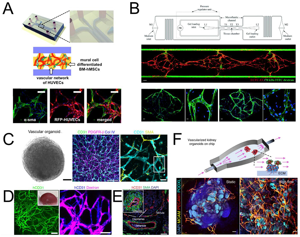
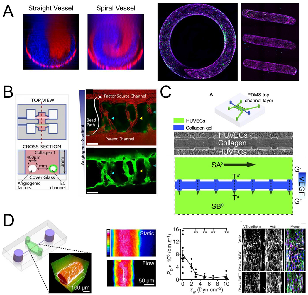
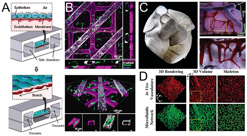
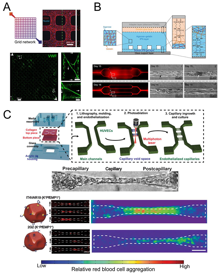
Organ-on-a-chip (OOC) platforms involve the miniaturization of cell culture systems and enable a variety of novel experimental approaches. These range from modeling the independent effects of biophysical forces on cells to screening novel drugs in multi-organ microphysiological systems, all within microscale devices. As in living systems, the incorporation of vascular structure is a key feature common to almost all organ-on-a-chip systems. In this review we highlight recent advances in organ-on-a-chip technologies with a focus on the vasculature. We first present the developmental process of the blood vessels through which vascular cells assemble into networks and remodel to form complex vascular beds under flow. We then review self-assembled vascular models and flow systems for the study of vascular development and biology as well as pre-patterned vascular models for the generation of perfusable microvessels for modeling vascular and tissue function. We finally conclude with a perspective on developing future OOC approaches for studying different aspects of vascular biology. We highlight the fit for purpose selection of OOC models towards either simple but powerful testbeds for therapeutic development, or complex vasculature to accurately replicate human physiology for specific disease modeling and tissue regeneration.

Keywords: Endothelial cells; Mechanotransduction; Microfluidics; Organ-on-a-chip; Organoids; Vascular biology.


Figures

Fig. 1.
Fig. 1.
Models of vascular self-assembly. (A) Endothelial cells and mural cells embedded in a fibrin matrix and supported by perfusion self-assemble into perfusable networks with close contact between cell types (adapted from Jeon et al[49]). (B) Controlled interstitial flow and mass transport in a microfluidic design (top) across EC-laden hydrogels stimulates vasculogenesis and the formation of perfusable vascular networks (adapted from Phan et al[51]). Middle panel: three tissue chamber with red for ECs and green for perfusion of 70kDa FITC-Dextran. Bottom panels: staining of claudin-5 (left two), and VE-Cad (right two). (C) Vascular organoids after 15 days of differentiation compact into a sphere comprised of CD31 expressing endothelial cells that are enveloped with mural cells (PDGFR-β, SMA) and basement membrane (COL IV). (D) hVOs transplanted in mice survive and form robust connection with the host vasculature. (E) > 1 month after transplantation, hVOs develop hierarchical vascular structures identifiable as arteries, venules, and capillaries (C,D,E adapted from Wimmer et al[62]). (F) Using microfluidics to deliver flow through vascularized kidney organoids enhanced vascular network formation as well as maturation of kidney organoid parenchyma (adapted from Homan et al[68]).
Fig. 2.
Fig. 2.
Engineering biophysical and biochemical cues in OOCs. (A) The flow in curved geometries is characterized by secondary flows and mixing not seen in straight vessels (left) and its effect can be studied in endothelialized spiral vessels (right, VECAD, magenta; VWF, green, adapted from Mandrycky et al[78]). (B) Endothelial cells cultured in channels can be stimulated by engineered biochemical gradients to stimulate angiogenesis and anastomosis to the factor source channel (adapted from Nguyen et al[86]). (C) Biochemical gradients can be combined with defined biophysical forces in opposing channels to investigate the influence of shear stress and interstitial flow differences on sprouting into collagen hydrogels (adapted from Song and Munn[88]). (D) Perfusion vascularized channels leads to a decrease in vascular permeability with increasing shear stress that can be disrupted using the γ-secretase inhibitor DAPT (adapted from Polacheck et al[90]).
Fig. 3.
Fig. 3.
Strategies for vascularization of multicellular organ-on-a-chip platforms: (A) A schematic for microfluidic-based approach to a vascularized lung-on-a-chip (adapted from Huh et al[100]) (B) A human renal tubule on a chip created through soft lithography showing tubular channels overlying endothelial channels with flanking orthogonal views (top) and perspective views showing perivascular association with the vessel (bottom, adapted from Rayner et al[109]). (C) A 3D human heart model bioprinted in collagen (left) with vessels perfused in red (top right) and highlighted area shown magnified (bottom right, adapted from Lee et al[113]). (D) In vivo vasculature (top) was used to create a photomask for direct laser-writing of channels into a PEG-based hydrogel (bottom, adapted from Heintz et al[114]).
Fig. 4.
Fig. 4.
Blood-endothelial interactions in organ-on-a-chip systems. (A) Lithographically patterned endothelialized grid networks release VWF (green) when stimulated, which form complex fiber meshes under perfusion (adapted from Zheng et al[119]). (B) High concentrations of irreversibly sickled red blood cells perfused through patterned microvascular networks in agarose-gelatin hydrogels cause occlusions and increase vascular permeability (“Day 15”) that can be recovered within 24 hours with continuous perfusion (adapted from Qiu et al[116]). (C) Generation of capillary scale vessels (5 – 10 pm diameter) is possible using a combined molding and photoablation strategy (top). Malaria infected red blood cells perfused through capillaries can aggregate and different parasite variants aggregate in different regions of the capillary space (adapted from Arakawa et al[115]).

References

    1. Burggren WW, Cardiovascular Development and Angiogenesis in the Early Vertebrate Embryo, Cardiovasc. Eng. Technol 4 (2013) 234–245. 10.1007/s13239-013-0118-x. - DOI - PubMed
    1. Patel-Hett S, D’Amore PA, Signal Transduction in Vasculogenesis and Developmental Angiogenesis, Int. J. Dev. Biol. 55 (2011) 353–363. 10.1387/ijdb.103213sp. - DOI - PMC - PubMed
    1. Heinke J, Patterson C, Moser M, Life is a pattern: vascular assembly within the embryo, Front. Biosci. Elite Ed. 4 (2012) 2269–2288. - PMC - PubMed
    1. Culver JC, Dickinson ME, The Effects of Hemodynamic Force on Embryonic Development, Microcirculation. 17 (2010) 164–178. 10.1111/j.1549-8719.2010.00025.x. - DOI - PMC - PubMed
    1. Han Y, Huang K, Yao Q-P, Jiang Z-L, Mechanobiology in vascular remodeling, Natl. Sci. Rev. 5 (2018) 933–946. 10.1093/nsr/nwx153. - DOI

Publication types