

Harnessing biomaterials for lymphatic system modulation
- PMID: 34118451
- PMCID: PMC9113193
- DOI: 10.1016/j.actbio.2021.06.006
Harnessing biomaterials for lymphatic system modulation
Abstract
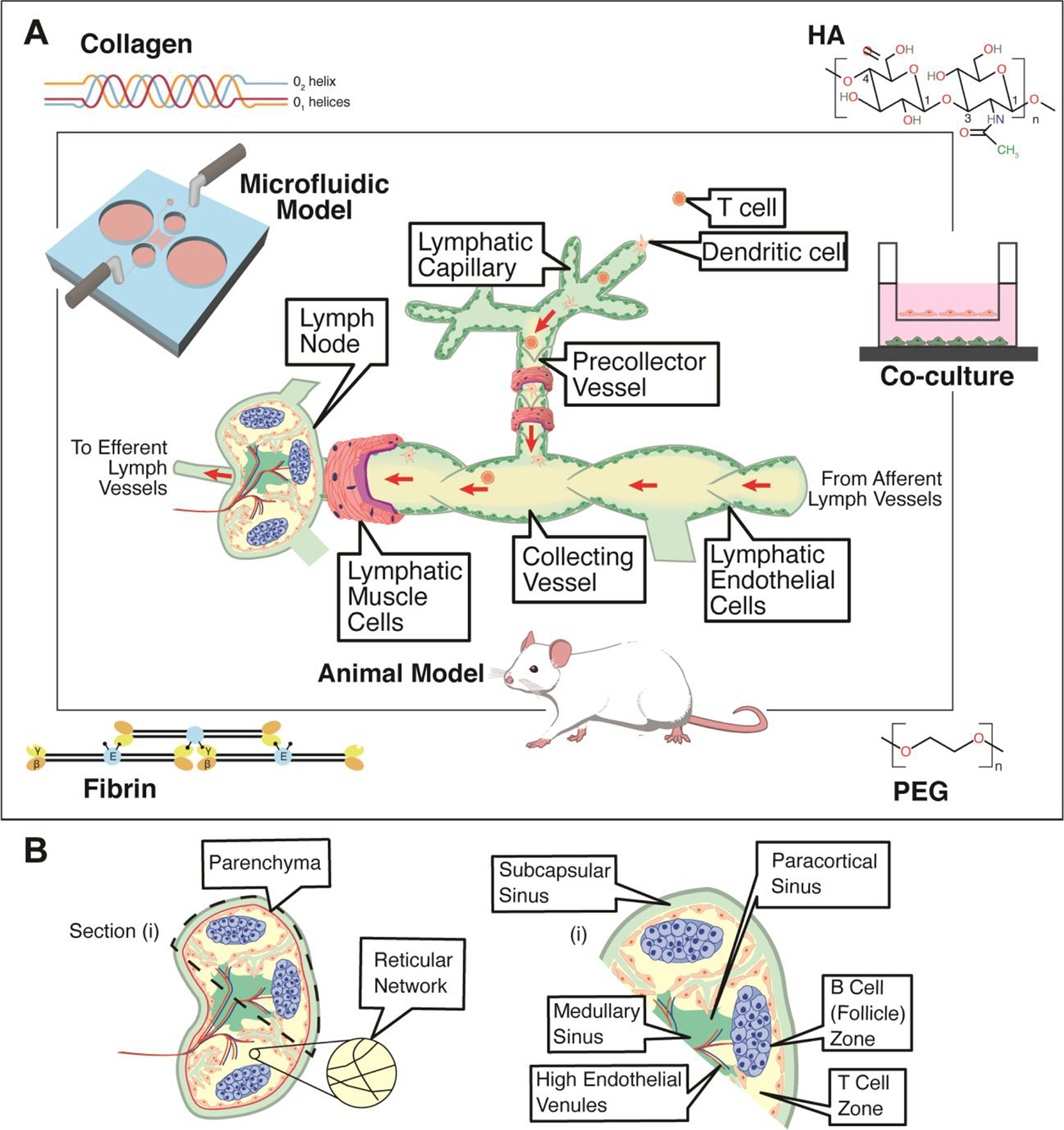
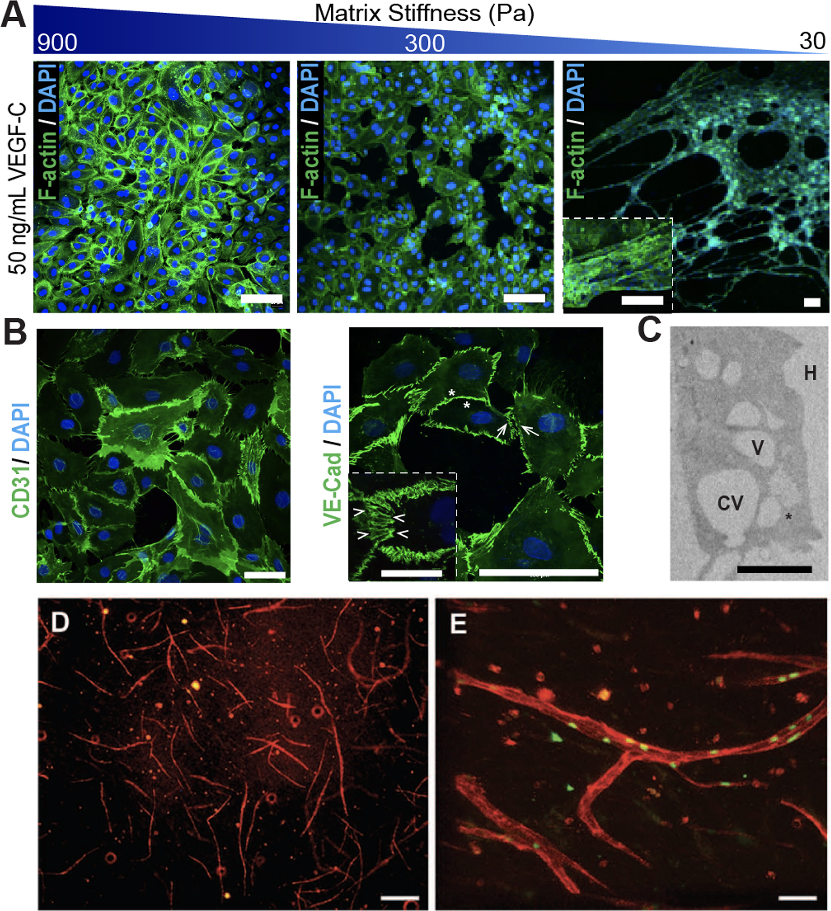
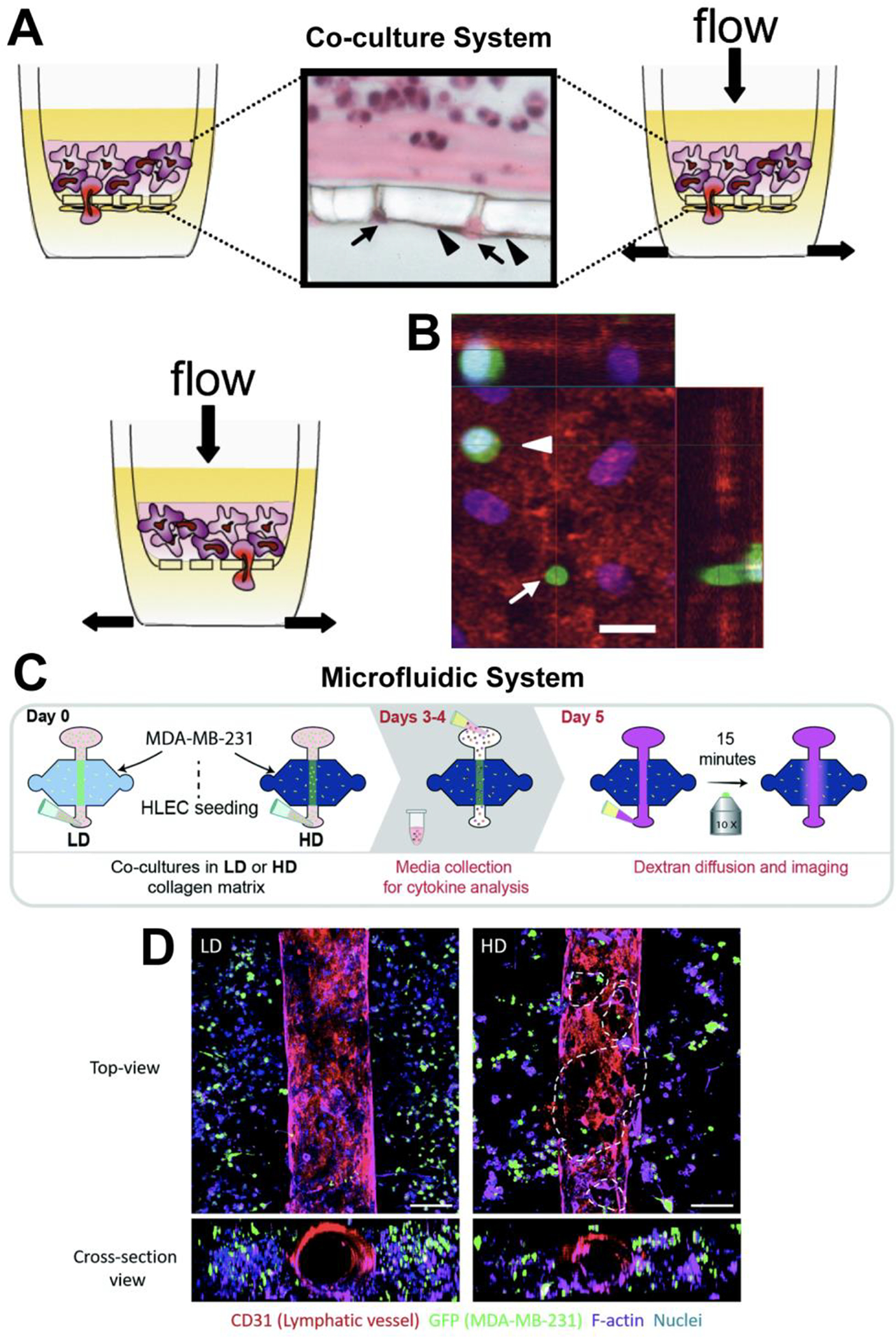
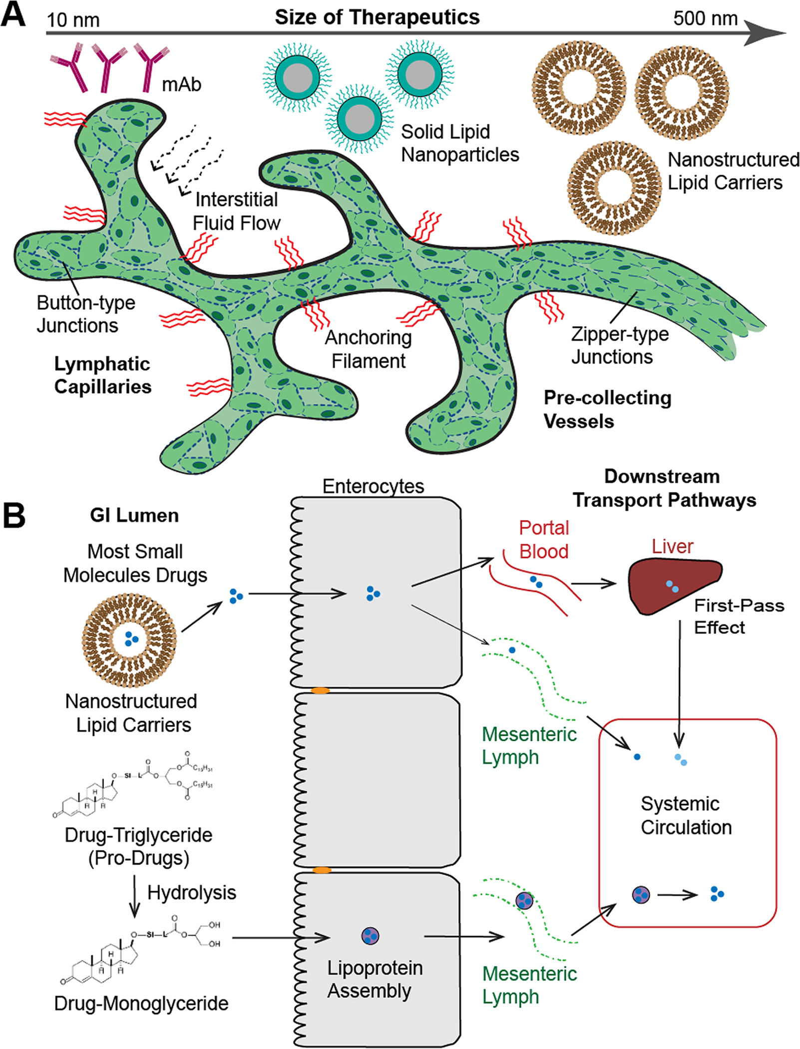
The lymphatic system plays an integral part in regulating immune cell trafficking and the transport of macromolecules. However, its influence on disease progression and drug uptake is understood less than that of the vascular system. To bridge this knowledge gap, biomaterials can be used to investigate the lymphatic system and to provide novel understanding into complex disease processes, including cancer metastasis and inflammation. Insight gained from these mechanistic studies can be further used to design innovative biomaterials to modulate the immune system, improve drug delivery, and promote tissue regeneration. This review article focuses on recent advances in (i) biomaterials used for lymphatic vessel formation, (ii) models for studying lymphatic-immune cells interactions, (iii) pharmaceuticals and their interactions with the lymphatic system, (iv) and strategies for drug delivery via the lymphatic system. Finally, several challenges regarding adopting biomaterials for immunomodulation and future perspectives are discussed. STATEMENT OF SIGNIFICANCE: The lymphatic system plays an integral part in regulating immune cell trafficking and the transport of macromolecules. However, its influence on disease progression and drug uptake is understood less than that of the vascular system. This review article focuses on recent progresses in biomaterials to investigate the lymphatic system and to provide novel understanding into complex disease states. Insight gained from these mechanistic studies can be further used to design innovative biomaterials to modulate the immune system, improve drug delivery, and promote tissue regeneration. Finally, a number of challenges in adopting biomaterials for immunomodulation and future perspectives are discussed.
Keywords: Biomaterials; Drug delivery; Immune system; Lymphatic vessels; Tissue engineering.
Copyright © 2021. Published by Elsevier Ltd.
Conflict of interest statement
Declaration of Competing Interest The authors declare that they have no known competing financial interests or personal relationships that could have appeared to influence the work reported in this paper.
Figures
References
Publication types
MeSH terms
Substances
Grants and funding
LinkOut - more resources
Full Text Sources
Research Materials
Miscellaneous
