. 2021 Jun:83:106015.
doi: 10.1016/j.ijscr.2021.106015. Epub 2021 May 26.

Emergency endoscopic surgery for pituitary apoplexy presenting as cerebral infarction in a limited resources condition: A case report

Affiliations

Emergency endoscopic surgery for pituitary apoplexy presenting as cerebral infarction in a limited resources condition: A case report

He Van Dong et al. Int J Surg Case Rep. 2021 Jun.

Abstract

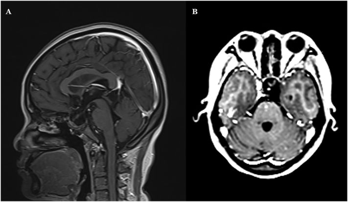
Introduction and importance: Pituitary apoplexy is defined as a sudden onset of neurologic deficit due to infarction or hemorrhage of the pituitary tumor. We report a case of emergency endoscopic surgery for pituitary apoplexy presenting as cerebral infarction due to ICA compression in a limited resources condition.

Case presentation: A 38-year-old female presented with acute onset of severe headache, decreased level of consciousness, decreased visual acuity bilaterally, aphasia, and right hemiparesis. Computed tomography angiography showed a hyperdense sellar mass with stenosis of the left ICA. The patient underwent emergent endoscopic transsphenoidal surgery for sellar decompression.

Clinical discussion: The epidermiology, presentation and diagnosis and strategy of treatments as well as their outcomes were discussed.

Conclusion: Pituitary apoplexy should be taken into consideration in a patient with increasing headache and neuro-ophthalmic symptoms. Pituitary apoplexy presenting as cerebral infarction is rare. The aim of surgery in emergency setting was sellar decompression. Endoscopic transsphenoidal surgery was an effective treatment.

Keywords: Cerebral infarction; Endoscopic transsphenoidal surgery; Pituitary adenoma; Pituitary apoplexy.


Conflict of interest statement

The authors declared no conflict of interest.

Figures

Fig. 1
Fig. 1
MRI image at the local hospital 1 day before the operation. A: The axial T1-weighted image with gadolinium showed a pituitary tumor with left lateral extension. B: The coronal T1-weighted image with gadolinium showed a pituitary tumor with upper extension.
Fig. 2
Fig. 2
The computed topography angiography imaging before the operation. 1: The right internal carotid artery. 2: The left internal carotid artery. 3: Pituitary tumor. A: Axial image with marked tumor. Notice the compression by the tumor of the clinoid and supraclinoid segments (C5-C6 segments) of the left ICA as its comparison to the right ICA. B: Sagittal image showed the compression by the tumor of the clinoid and supraclinoid segments (C5–C6 segments) of the left ICA. C: 3-D reconstruction image of the cerebral artery system. D: Coronal image with marked tumor. Notice the compression by the tumor of the clinoid and supraclinoid segments (C5–C6 segments) of the left ICA as its comparison to the right ICA. E: Axial image showed hypoattenuation in the left hemisphere. Notes in Fig. 2: The compression of the left ICA by the tumor; the diameter of the left ICA in comparison with the right ICA; no thrombus was found in the cerebral arteries.
Fig. 3
Fig. 3
Intraoperative endoscopic images. A: The pituitary tumor. Notice the infarction inside the tumor. B: The sellar diaphragm. No residual tumor was observed.
Fig. 4
Fig. 4
Microscopic image of the tumor in H&E stain in 40×. Notice the mass infarction of the adenoma cells.
Fig. 5
Fig. 5
The CT scan at 5 days post operation. A: No hemorrhage in the sellar region was observed. B: The hemorrhage inside the infarction region of the left hemisphere.
Fig. 6
Fig. 6
10 days post operation MRI images. A: T1 with gadolinium images shows no residual tumor. B: DWI image showed the left hemisphere infarction. C: 3D-TOF image showed the left ICA stenosis.
Fig. 7
Fig. 7
4 months post operation. A, B: T1 with gadolinium images shows no residual tumor.

References

    1. Verrees M., Arafah B.M., Selman W.R. Pituitary tumor apoplexy: characteristics, treatment, and outcomes. Neurosurg. Focus. Apr 15 2004;16(4):E6. doi: 10.3171/foc.2004.16.4.7. - DOI - PubMed
    1. A D., Ronquillo Yasmyne. Snellen Chart. Text. 2020. https://www.ncbi.nlm.nih.gov/books/NBK558961/ 2020/06/10.
    1. Kniestedt C., Stamper R.L. Visual acuity and its measurement. Ophthalmol. Clin. N. Am. Jun 2003;16(2):155–170. doi: 10.1016/s0896-1549(03)00013-0. v. - DOI - PubMed
    1. Rajasekaran S., Vanderpump M., Baldeweg S. UK guidelines for the management of pituitary apoplexy. Clin. Endocrinol. Jan 2011;74(1):9–20. doi: 10.1111/j.1365-2265.2010.03913.x. - DOI - PubMed
    1. Jho D.H., Biller B.M., Agarwalla P.K., Swearingen B. Pituitary apoplexy: large surgical series with grading system. World Neurosurg. Nov 2014;82(5):781–790. doi: 10.1016/j.wneu.2014.06.005. - DOI - PubMed