. 2021 Jun 12;16(1):104.
doi: 10.1186/s13014-021-01829-y.

Impact of superparamagnetic iron oxide nanoparticles on in vitro and in vivo radiosensitisation of cancer cells

Affiliations

Impact of superparamagnetic iron oxide nanoparticles on in vitro and in vivo radiosensitisation of cancer cells

Emily Russell et al. Radiat Oncol. .

Abstract

Purpose: The recent implementation of MR-Linacs has highlighted theranostic opportunities of contrast agents in both imaging and radiotherapy. There is a lack of data exploring the potential of superparamagnetic iron oxide nanoparticles (SPIONs) as radiosensitisers. Through preclinical 225 kVp exposures, this study aimed to characterise the uptake and radiobiological effects of SPIONs in tumour cell models in vitro and to provide proof-of-principle application in a xenograft tumour model.

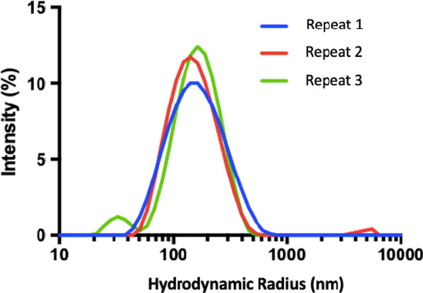
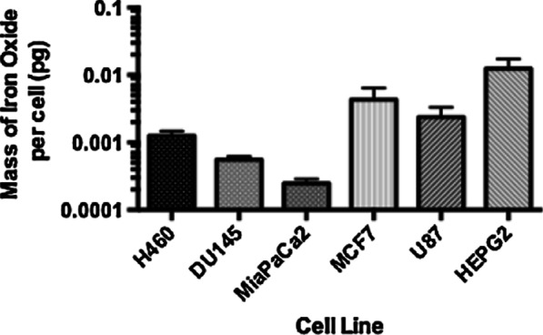
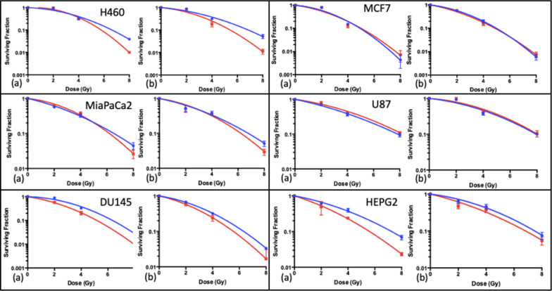
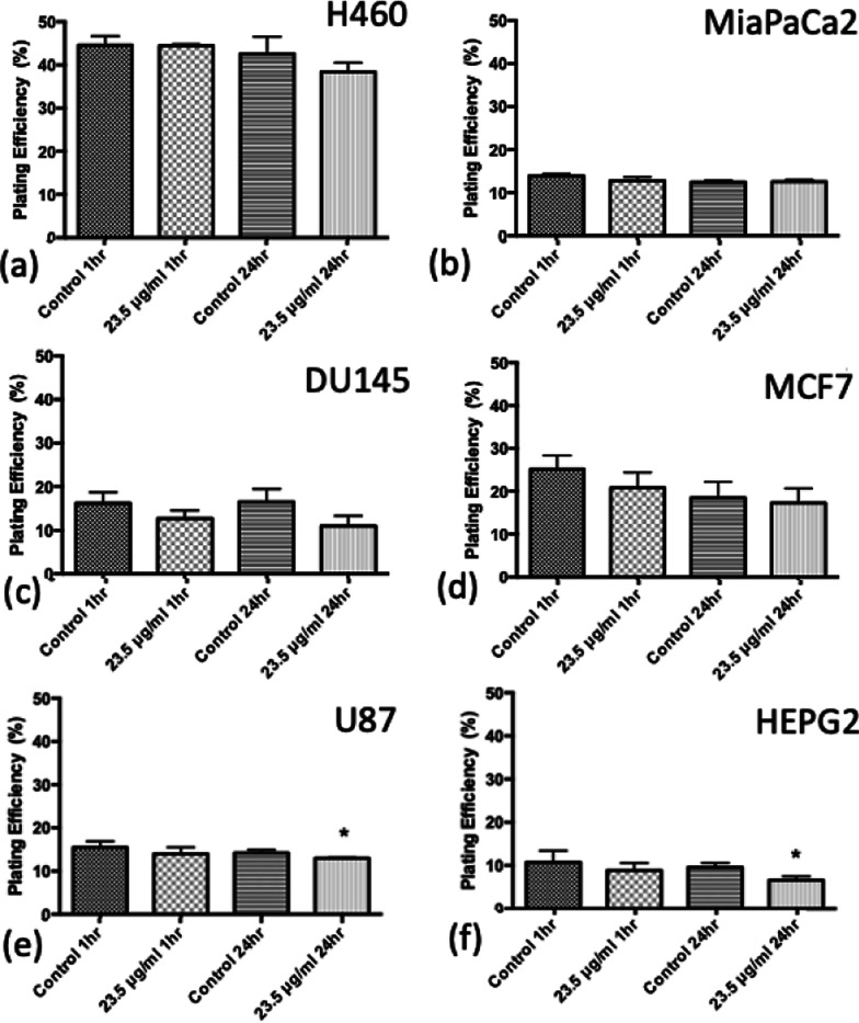
Methods: SPIONs were also characterised to determine their hydrodynamic radius using dynamic light scattering and uptake was measured using ICP-MS in 6 cancer cell lines; H460, MiaPaCa2, DU145, MCF7, U87 and HEPG2. The impact of SPIONs on radiobiological response was determined by measuring DNA damage using 53BP1 immunofluorescence and cell survival. Sensitisation Enhancement Ratios (SERs) were compared with the predicted Dose Enhancement Ratios (DEFs) based on physical absorption estimations. In vivo efficacy was demonstrated using a subcutaneous H460 xenograft tumour model in SCID mice by following intra-tumoural injection of SPIONs.

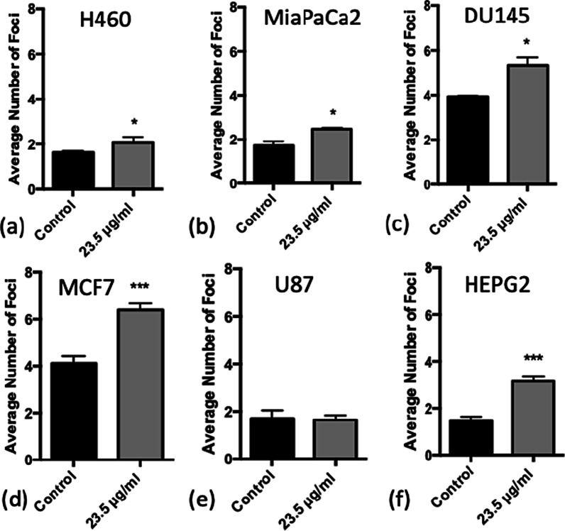
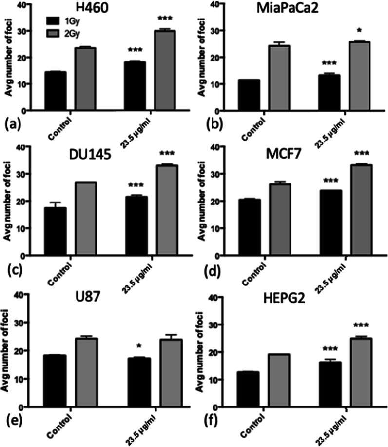
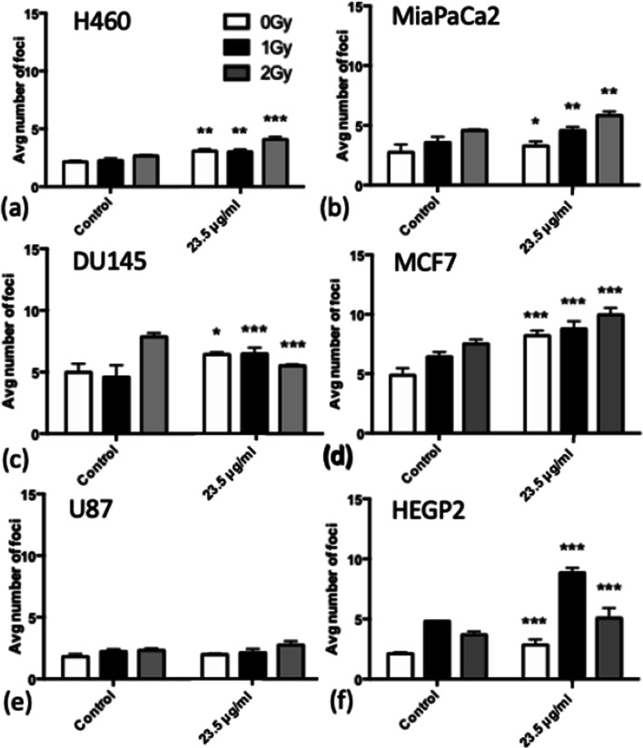
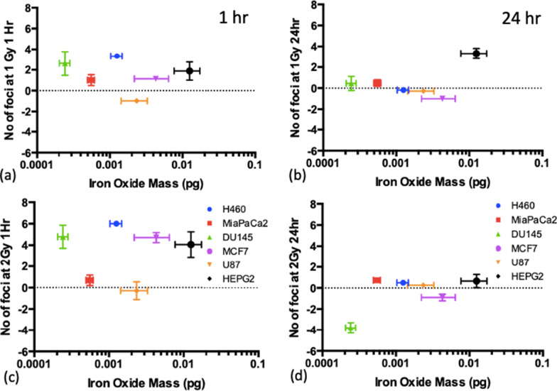
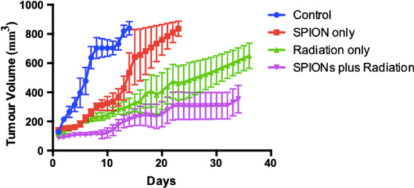
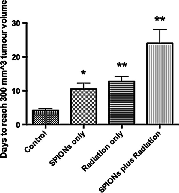
Results: The hydrodynamic radius was found to be between 110 and 130 nm, with evidence of being monodisperse in nature. SPIONs significantly increased DNA damage in all cell lines with the exception of U87 cells at a dose of 1 Gy, 1 h post-irradiation. Levels of DNA damage correlated with the cell survival, in which all cell lines except U87 cells showed an increased sensitivity (P < 0.05) in the linear quadratic curve fit for 1 h exposure to 23.5 μg/ml SPIONs. There was also a 30.1% increase in the number of DNA damage foci found for HEPG2 cells at 2 Gy. No strong correlation was found between SPION uptake and DNA damage at any dose, yet the biological consequences of SPIONs on radiosensitisation were found to be much greater, with SERs up to 1.28 ± 0.03, compared with predicted physical dose enhancement levels of 1.0001. In vivo, intra-tumoural injection of SPIONs combined with radiation showed significant tumour growth delay compared to animals treated with radiation or SPIONs alone (P < 0.05).

Conclusions: SPIONs showed radiosensitising effects in 5 out of 6 cancer cell lines. No correlation was found between the cell-specific uptake of SPIONs into the cells and DNA damage levels. The in vivo study found a significant decrease in the tumour growth rate.

Keywords: Nanoparticles; Radiobiology.


Conflict of interest statement

The authors declare that they have no competing interests.

Figures

Fig. 1
Fig. 1
DLS measurements indicating distribution of hydrodynamic radius of SPIONs, at the stock concentration which is the concentration that the supplier provides; 5 mg/ml
Fig. 2
Fig. 2
Uptake measurements performed using ICP-MS, representing the mass of iron oxide taken up per cell by each cell lines, for an added concentration of 23.5 μg/ml and an incubation time of 24 h, presented as mean ± SEM. (n = 3)
Fig. 3
Fig. 3
Average number of DNA damage foci counted for each cell line when treated with 23.5 μg/ml SPIONs in the absence of radiation, with 24 h incubation. a H460, b MiaPaCa2, c DU145, d MCF7, e U87 and f HEPG2. (n = 3) Presented as mean ± SD with statistical significance represented as; P < 0.05; *; P < 0.001: **; P < 0.0001: ***
Fig. 4
Fig. 4
Average number of DNA damage foci counted for cells treated with 23.5 μg/ml SPIONs 1 h after exposure to 1 Gy or 2 Gy of X-rays. a H460, b MiaPaCa2, c DU145, d MCF7, e U87 and f HEPG2. (n = 3) Presented as mean ± SD with statistics represented as; P < 0.05: *; P < 0.001: **; P < 0.0001: ***
Fig. 5
Fig. 5
Average number of DNA damage foci counted for each cell line when treated with 23.5 μg/ml SPIONs and doses of 0–2 Gy measured at 24 h after irradiation. a H460, b MiaPaCa2, c DU145, d MCF7, e U87 and f HEPG2. (n = 3) Presented as mean ± SD with statistics represented as; P < 0.05: *; P < 0.001: **; P < 0.0001: ***
Fig. 6
Fig. 6
Mass of iron oxide detected per cell for each cell line compared to the number of foci counted, corrected for controls, and also corrected for the number of foci for cells without SPIONs, at the 1 h timepoint (left) and 24 h timepoint (right) after combined exposure with SPIONs and 225 kVp X-rays for; a, c 1 Gy and b, d 2 Gy. Presented as mean ± SD. Spearman Correlation coefficient, ρ, is indicated on each graph
Fig. 7
Fig. 7
Clonogenic cell survival curves fitted with the linear quadratic model for the combination of 225 kVp X-rays with 23.5 μg/ml SPIONs, for a 1 h exposure time; a 1 h and b 24 h exposure times. (n = 3) Presented as mean ± SD
Fig. 8
Fig. 8
Plating efficiency for cell lines in the absence of radiation, for control samples and 23.5 μg/ml SPIONs for both 1 h and 24 h exposure times. a H460, b MiaPaCa2, c DU145, d MCF7, e U87 and f HEPG2. (n = 3) Presented as mean ± SD with statistics represented as; P < 0.05: *; P < 0.001: **; P < 0.0001: ***
Fig. 9
Fig. 9
Tumour Volume with time for in vivo experiment for 4 subgroups; control, SPION only, radiation only, and SPIONs plus radiation, taken as the median value across all mice, using linear interpolation for days between measurements. Presented as mean ± SEM
Fig. 10
Fig. 10
Average time (days) for tumour volume to reach 300 mm3 for the 4 subgroups; control, SPIONs only, radiation only, and SPIONs plus radiation. Presented as mean ± SEM with statistics represented as; P < 0.05: *; P < 0.001: **; P < 0.0001: ***

References

    1. Bekelman JE, Lee WR. Six questions to ask before we shorten radiation treatments for intact prostate cancer. Int J Radiat Oncol Biol Phys. 2017;97(4):718–721. doi: 10.1016/j.ijrobp.2016.11.038. - DOI - PubMed
    1. Lee WR. Hypofractionation for prostate cancer: tested and proven. Lancet Oncol. 2016;17(8):1020–1022. doi: 10.1016/S1470-2045(16)30150-4. - DOI - PubMed
    1. Symonds P, Jones GDD. FLASH radiotherapy: The next technological advance in radiation therapy? Clin Oncol. 2019;31(7):405–406. doi: 10.1016/j.clon.2019.05.011. - DOI - PubMed
    1. Bourhis J, Montay-Gruel P, Gonçalves Jorge P, Bailat C, Petit B, Ollivier J, et al. Clinical translation of FLASH radiotherapy: Why and how? Radiother Oncol. 2019;139:11–17. doi: 10.1016/j.radonc.2019.04.008. - DOI - PubMed
    1. Vozenin MC, Hendry JH, Limoli CL. Biological benefits of ultra-high dose rate FLASH radiotherapy: sleeping beauty awoken. Clin Oncol. 2019;31(7):407–415. doi: 10.1016/j.clon.2019.04.001. - DOI - PMC - PubMed

Substances