. 2021 Jun;44(3):293-303.
doi: 10.1016/j.bj.2021.01.005. Epub 2021 Jan 28.

Perilla (Perilla frutescens) leaf extract inhibits SARS-CoV-2 via direct virus inactivation

Affiliations

Perilla (Perilla frutescens) leaf extract inhibits SARS-CoV-2 via direct virus inactivation

Wen-Fang Tang et al. Biomed J. 2021 Jun.

Abstract

Background: While severe acute respiratory syndrome coronavirus 2 (SARS-CoV-2) infection presents with mild or no symptoms in most cases, a significant number of patients become critically ill. Remdesivir has been approved for the treatment of coronavirus disease 2019 (COVID-19) in several countries, but its use as monotherapy has not substantially lowered mortality rates. Because agents from traditional Chinese medicine (TCM) have been successfully utilized to treat pandemic and endemic diseases, we designed the current study to identify novel anti-SARS-CoV-2 agents from TCM.

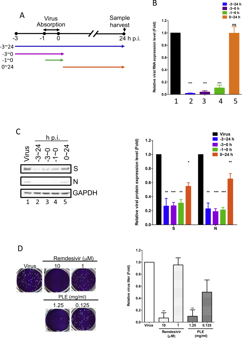
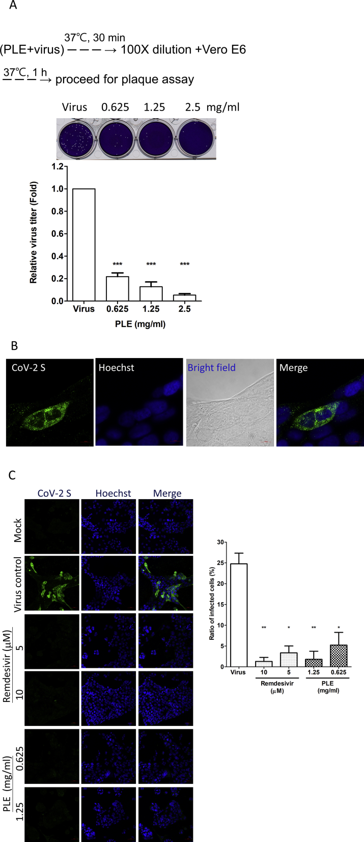
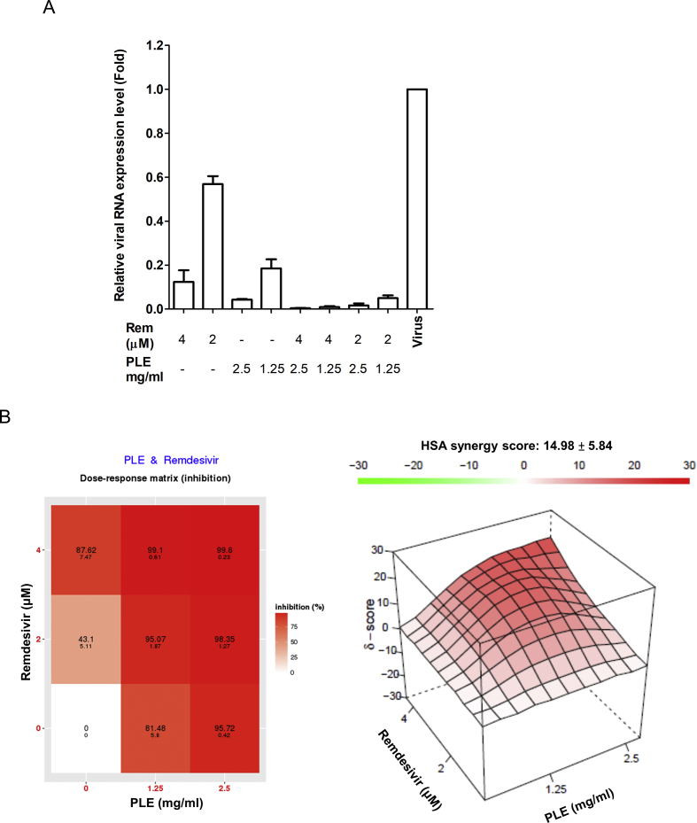
Methods: We initially used an antivirus-induced cell death assay to screen a panel of herbal extracts. The inhibition of the viral infection step was investigated through a time-of-drug-addition assay, whereas a plaque reduction assay was carried out to validate the antiviral activity. Direct interaction of the candidate TCM compound with viral particles was assessed using a viral inactivation assay. Finally, the potential synergistic efficacy of remdesivir and the TCM compound was examined with a combination assay.

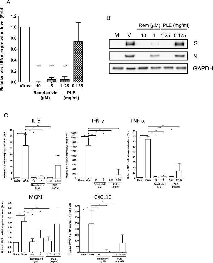
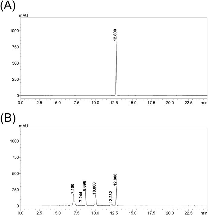
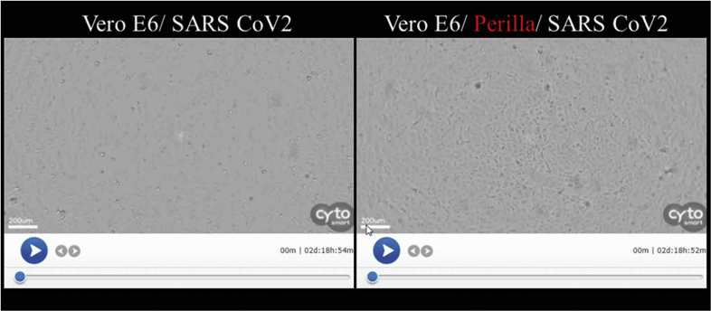
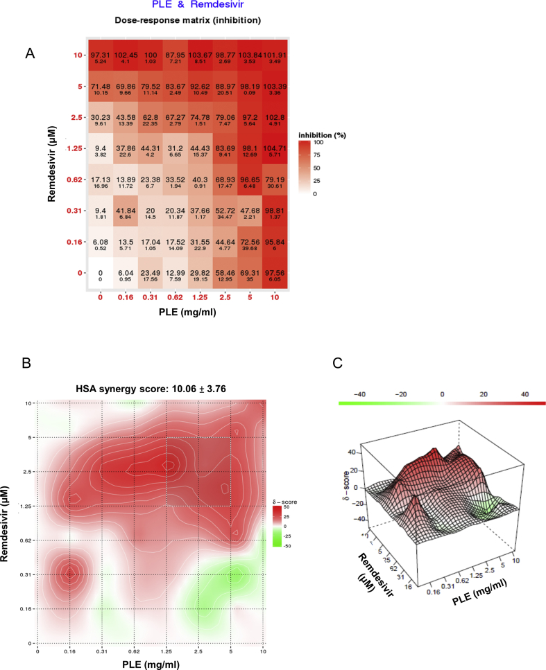
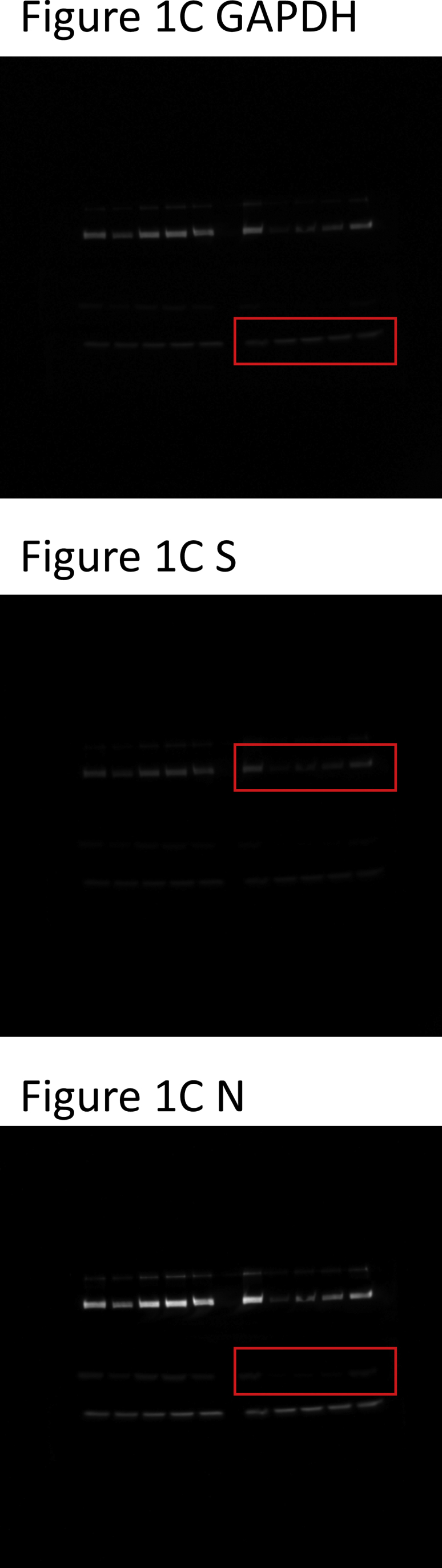
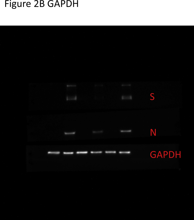
Results: The herbal medicine Perilla leaf extract (PLE, approval number 022427 issued by the Ministry of Health and Welfare, Taiwan) had EC50 of 0.12 ± 0.06 mg/mL against SARS-CoV-2 in Vero E6 cells - with a selectivity index of 40.65. Non-cytotoxic PLE concentrations were capable of blocking viral RNA and protein synthesis. In addition, they significantly decreased virus-induced cytokine release and viral protein/RNA levels in the human lung epithelial cell line Calu-3. PLE inhibited viral replication by inactivating the virion and showed additive-to-synergistic efficacy against SARS-CoV-2 when used in combination with remdesivir.

Conclusion: Our results demonstrate for the first time that PLE is capable of inhibiting SARS-CoV-2 replication by inactivating the virion. Our data may prompt additional investigation on the clinical usefulness of PLE for preventing or treating COVID-19.

Keywords: COVID-19; Coronavirus; Perilla frutescens (L.) Britt; SARS-CoV-2; Traditional Chinese medicine; Zisu.


Conflict of interest statement

Conflicts of interest The authors declare they have no actual or potential competing financial interests.

Figures

Fig. 1
Fig. 1
Treatment with PLE inhibits SARS-CoV-2 at early stages of replication. (A) Schematic representation of the time-of-addition assay. (B–C) Vero E6 cells were infected with SARS-CoV-2 at a MOI of 0.01. Subsequently, PLE (1.25 mg/mL) was added at the following time points: before virus entry (between −3 and 0 h p.i.), during virus absorption (−1−0 h p.i.), and following virus adsorption (0–24 h p.i.). Infected cells were collectively harvested at 24 h p.i.; viral RNA synthesis and viral protein expression were analyzed with qPCR (B) and western blotting (C), respectively. (B) Expression levels of viral RNA were initially normalized to GAPDH mRNA at each experimental condition. Moreover, the ratio measured in PLE-treated cells was normalized to the RNA level of virus control (arbitrarily set to 1). (C) The intensity of SARS-CoV-2 spike protein (S) and nucleocapsid (N) expression was normalized to GAPDH. Moreover, the ratio measured in PLE-treated cells was normalized to the protein level of virus control (arbitrarily set to 1). N = 3. (D) The results of the plaque reduction assay revealed that SARS-CoV-2 infectivity was diminished after exposure of Vero E6 cells to PLE. SARS-CoV-2 was pre-incubated with various concentrations of PLE or remdesivir before its addition to Vero E6 cells for the plaque assay. The number of plaques was calculated and normalized to that of virus control (arbitrarily set to 1). Data in bar charts are expressed as means ± standard error of the mean from at least two independent experiments. ∗p < 0.05, ∗∗p < 0.01, and ∗∗∗p < 0.005; ns = not significant.
Fig. 2
Fig. 2
PLE inhibits SARS-CoV-2 replication in Calu-3 cells. The expression of viral RNA and proteins (A–B) and cytokine mRNA (C) in Calu-3 cells was inhibited by PLE. Calu-3 cells were infected with SARS-CoV-2 in presence of various concentrations of PLE with remdesivir serving as positive control. Upon cell harvesting, RNA and viral protein quantification was performed with qRT-PCR (A) and western blotting (B), respectively. (A, C) Expression levels of viral or cytokine RNA were initially normalized to GAPDH mRNA. Moreover, the ratio measured in PLE-treated cells was normalized to the RNA level of virus control (arbitrarily set to 1). Data are expressed as means ± standard error of the mean from at least three independent experiments. ∗p < 0.05, ∗∗p < 0.01, and ∗∗∗p < 0.005.
Fig. 3
Fig. 3
SARS-CoV-2 was inactivated by PLE. (A) The virus stock was pre-treated with increasing concentrations of PLE, and the remaining viral titers were subsequently determined using a plaque assay carried out in Vero E6 cells. The number of plaques for PLE-treated viruses was normalized to that of the virus control (arbitrarily set to 1). Data are expressed as means ± standard error of the mean from at least three independent experiments. (B–C) Confocal immunofluorescence microscopy revealed that PLE treatment reduced viral protein synthesis in Calu-3 cells. Cells treated with or without remdesivir and PLE were infected with SARS-CoV-2 at a MOI of 0.01. Cells were harvested at 48 h p.i. for confocal microscopy using the anti-S antibodies as indicated. Fluorescence images of S protein subcellular distribution in SARS-CoV-2-infected Calu-3 cells obtained either in absence (B) or presence (C) of inhibitors. Magnifications of objective lenses: 100 × (B) and 20 × (C). Nuclei were stained with a Hoechst dye. The transmitted light in the bright field revealed the overall morphology of Calu-3 cells. The bar chart in the right panel illustrates the ratios of infected cells under different experimental conditions (magnification of objective lens: 20 × ). For each condition, the ratio of spike-positive cells was calculated in two independent experiments from >200 cells in randomly selected fields. ∗p < 0.05, ∗∗p < 0.01, and ∗∗∗p < 0.005.
Fig. 4
Fig. 4
Antiviral activity of PLE and remdesivir used in combination (A). Vero E6 cells were infected with SARS-CoV-2 with serial dilutions of PLE in combination with remdesivir and subsequently harvested at 24 h p.i. to quantify viral RNA loads by qRT-PCR. Expression levels of viral RNA were initially normalized to GAPDH mRNA. The ratio of drug-treated cells was subsequently normalized to the RNA level of virus control (arbitrarily set to 1). (B) The ratio of drug inhibition elicited by the combination of PLE and remdesivir was calculated with the HSA reference synergy model available in the SynergyFinder software (version 2). The graphs illustrate average results from two independent experiments carried out in duplicate.
figs1
figs1
Figure S1. PLE fingerprint analysis. PLE and rosmarinic acid were analyzed using a Shimadzu Nexera-I LC-2040C-3D HPLC system (Kyoto, Japan) with a Cosmosil 5C18-MS-II HPLC column (4.6 × 250 mm; Nacalai Tesque, Inc., Kyoto, Japan). PLE (50 mg/mL) and rosmarinic acid (5 μg/mL) were filtered through a 0.22-μm filter before analysis. Solvents consisted of 0.1% formic acid aqueous solution and acetonitrile. An acetonitrile gradient elution was carried out at the following conditions: 15−40%, 0−18 min; 40−100%, 18−20 min; 100%, 20−25 min, 100−15%, 25−35 min. The flow rate was 1 mL/min, whereas the detection wavelength and the injection volume were 330 nm and 20 μL, respectively. (A) Rosmarinic acid was detected at a retention time of 12.800 min; (B) Rosmarinic acid in PLE was detected at a retention time of 12.808 min.
figs2
figs2
Figure S2. Video clip of the cytopathic effect assay obtained using a real-time CytoSmart Lux2 imaging system. The filming was carried out over a 48-h timeframe. Vero-E6 cells were infected with SARS-CoV-2 at a MOI 0.01 either in the presence or absence of PLE.
figs3
figs3
Figure S3. Antiviral activity of PLE and remdesivir as observed in the combination assay. Vero E6 cells were infected with SARS-CoV-2 in the presence of serial dilutions of PLE combined with remdesivir. Cells were subsequently harvested at 72 h p.i. and cell viability was quantified with the MTT assay. The absorbance of drug-treated cells was normalized to mock-infected cells (arbitrarily set to 1). The ratio of drug inhibition elicited by the combination of PLE and remdesivir was calculated with the HSA reference synergy model available in the SynergyFinder software (version 2). The dose-response plot (A) as well as the two-dimensional (B) and three-dimensional (C) synergy maps are reported. Results should be considered representative of those obtained from two independent experiments carried out in duplicate.
figs4
figs4
figs5
figs5

References

    1. Coronaviridae Study Group of the International Committee on Taxonomy of V The species Severe acute respiratory syndrome-related coronavirus: classifying 2019-nCoV and naming it SARS-CoV-2. Nat Microbiol. 2020;5:536–544. - PMC - PubMed
    1. Hoffmann M., Kleine-Weber H., Schroeder S., Kruger N., Herrler T., Erichsen S. SARS-CoV-2 cell entry depends on ACE2 and TMPRSS2 and is blocked by a clinically proven protease inhibitor. Cell. 2020;181:271–280 e8. - PMC - PubMed
    1. Walls A.C., Park Y.J., Tortorici M.A., Wall A., McGuire A.T., Veesler D. Structure, function, and antigenicity of the SARS-CoV-2 spike glycoprotein. Cell. 2020;181:281–292 e6. - PMC - PubMed
    1. Masters P.S., Perlman S. Coronaviridae. In: David M., Knipe P.M.H., Martin Malcolm A., Griffin Diane E., Lamb Robert A., Bernard Roizman, Straus Stephen E., editors. Fields virology. Lippincott Williams & Wilkins; Philadelphia: 2013. pp. 825–858.
    1. Huang C., Wang Y., Li X., Ren L., Zhao J., Hu Y. Clinical features of patients infected with 2019 novel coronavirus in Wuhan, China. Lancet. 2020;395:497–506. - PMC - PubMed

LinkOut - more resources