. 2021 Aug:209:108663.
doi: 10.1016/j.exer.2021.108663. Epub 2021 Jun 11.

The aging mouse lens transcriptome

Affiliations

The aging mouse lens transcriptome

Adam P Faranda et al. Exp Eye Res. 2021 Aug.

Abstract

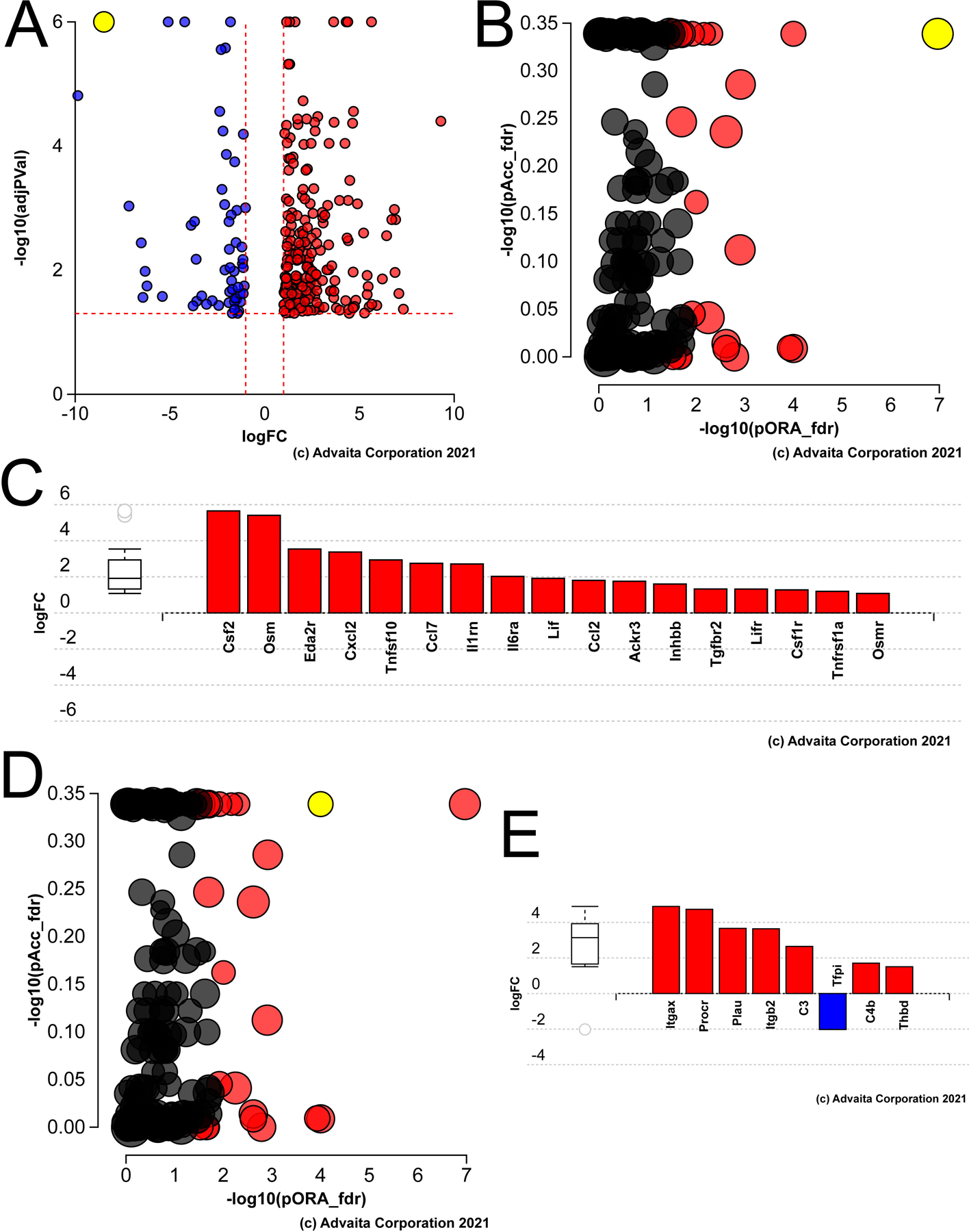
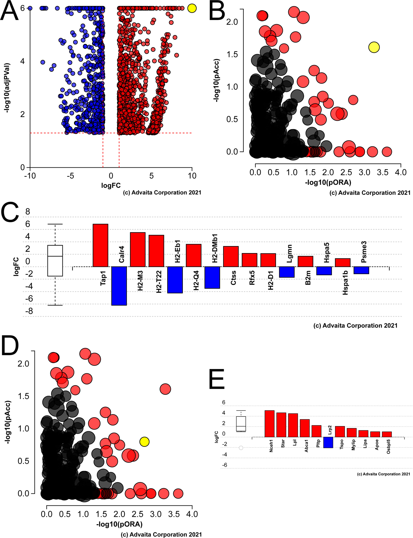
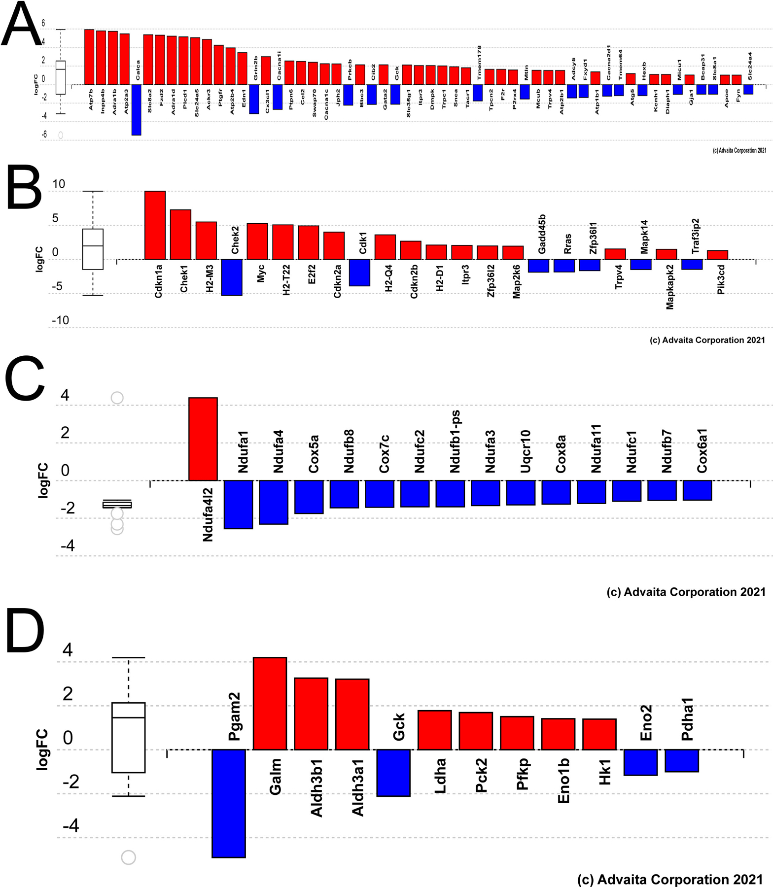
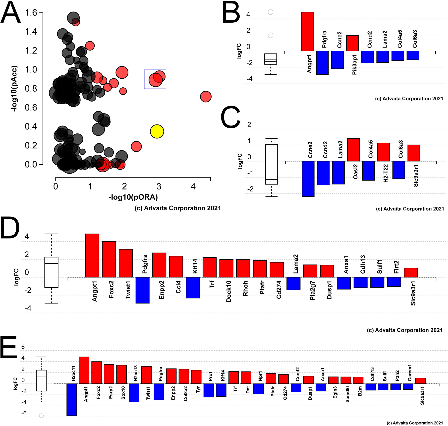
Age is a major risk factor for cataract (ARC). However, the influence of aging on the lens transcriptome is under studied. Lens epithelial (LEC) and fiber cells (LFC) were isolated from young (3 month old) and aged (24 month old) C57BL/6J mice, and the transcriptome elucidated via RNAseq. EdgeR estimated differential gene expression in pairwise contrasts, and Advaita's Ipathway guide and custom R scripts were used to evaluate the potential biological significance of differentially expressed genes (DEGs). This analysis revealed age-dependent decreases in lens differentiation marker expression in both LECs and LFCs, with gamma crystallin transcripts downregulating nearly 50 fold in aged LFCs. The expression of the transcription factors Hsf4 and Maf, which are known to activate lens fiber cell preferred genes, are downregulated, while FoxE3, which represses gamma crystallin expression, is upregulated in aged fibers. Aged LECs upregulate genes controlling the immune response, complement pathways, and cellular stress responses, including glutathione peroxidase 3 (Gpx3). Aged LFCs exhibit broad changes in the expression of genes regulating cell communication, and upregulate genes involved in antigen processing/presentation and cholesterol metabolism, while changes in the expression of mitochondrial respiratory chain genes are consistent with mitochondrial stress, including upregulation of NDufa4l2, which encodes an alternate electron transport chain protein. However, age did not profoundly affect the response of LECs to injury as both young and aged LECs upregulate inflammatory gene signatures at 24 h post injury to similar extents. These RNAseq profiles provide a rich data set that can be mined to understand the genetic regulation of lens aging and how this impinges on the pathophysiology of age related cataract.

Keywords: Aging; Cholestero; Complement pathways; Lens; Metabolism; Posterior capsular opacification; Senescence; Transcriptome.


Figures

Figure 1
Figure 1
Pathway analysis of genes differentially expressed in aged versus young mouse lens epithelial cells A) Volcano plot of the genes whose expression was statistically different between aged versus young LECs, yellow dot represents γF-crystallin B) Impact analysis of the DEGs suggest that the KEGG pathway map “cytokine-cytokine receptors” (yellow dot) is likely to be the most significantly impacted pathway in the aged lens epithelium. C) Bar graph showing the cytokine-cytokine receptor genes that are differentially expressed in the aged lens epithelium. D) Impact analysis showing that the second significant pathway in the aged lens epithelium represents genes involved in the complement pathway (yellow dot). E) Bar graph showing the complement pathway genes differentially expressed in the aged mouse lens epithelium.
Figure 2
Figure 2
Pathway analysis of genes differentially expressed in aged versus young mouse lens fiber cells A) Volcano plot of the genes whose expression was statistically different between aged versus young LECs, yellow dot represents cdkn1a (P21) B) Impact analysis of the DEGs suggest that the KEGG pathway map “antigen processing and presentation” (yellow dot) is likely to be the most significantly impacted pathway in the aged lens fibers. C) Bar graph showing the antigen processing and presentation genes that are differentially expressed in the aged lens fibers. D) Impact analysis showing that the another significantly impacted pathway in the aged lens fibers represents genes involved in cholesterol metabolism (yellow dot). E) Bar graph showing the cholesterol metabolism genes differentially expressed in the aged mouse lens fibers.
Figure 3
Figure 3
Genes differentially expressed in aged versus young mouse lens fiber cells grouped by gene ontology (GO) terms or Kegg pathways potentially relevant to age-related changes in lens biology A) Bar graph representing the DEGs in aged lens fibers mapping to the Kegg pathway cellular calcium homeostasis. B) Bar graph representing the DEGs in aged lens fibers mapping to the Kegg pathway cellular senescence. C) Bar graph representing the DEGs in aged lens fibers mapping to the GO term respiratory chain complex. D) Bar graph representing the DEGs in aged lens fibers mapping to the Kegg pathway glycolysis/gluconeogenesis.
Figure 4
Figure 4
Genes differentially expressed in aged versus young mouse lens epithelial cells at 24 hours PCS. A) Impact analysis of the 24 hour PCS DEGs with the yellow dot representing Akt signaling; boxed dots represent “human papilloma virus infection” and “alcoholism” B) Bar graph representing the DEGs in aged LECs at 24 hours PCS known to be involved in “Akt signaling”. C) Bar graph representing the DEGs in aged LECs at 24 hours PCS known to be involved in “human papilloma virus infection”. D) Bar graph representing the DEGs in aged LECs at 24 hours PCS mapped to the gene ontology term “cell motility”. D) Bar graph representing the DEGs in aged LECs at 24 hours PCS the gene ontology term “cell proliferation”.
Figure 5
Figure 5
Summary of the major effects of age on the mouse lens transcriptome

References

    1. Ackert-Bicknell CL, Anderson LC, Sheehan S, Hill WG, Chang B, Churchill GA, Chesler EJ, Korstanje R, Peters LL, 2015. Aging Research Using Mouse Models. Curr Protoc Mouse Biol 5, 95–133. - PMC - PubMed
    1. Aging Atlas C, 2021. Aging Atlas: a multi-omics database for aging biology. Nucleic Acids Res 49, D825–D830. - PMC - PubMed
    1. Agrawal SA, Anand D, Siddam AD, Kakrana A, Dash S, Scheiblin DA, Dang CA, Terrell AM, Waters SM, Singh A, Motohashi H, Yamamoto M, Lachke SA, 2015. Compound mouse mutants of bZIP transcription factors Mafg and Mafk reveal a regulatory network of non-crystallin genes associated with cataract. Hum Genet 134, 717–735. - PMC - PubMed
    1. Ahsan S, Draghici S, 2017. Identifying Significantly Impacted Pathways and Putative Mechanisms with iPathwayGuide. Curr Protoc Bioinformatics 57, 7 15 11–17 15 30. - PubMed
    1. Aleo MD, Doshna CM, Baltrukonis D, Fortner JH, Drupa CA, Navetta KA, Fritz CA, Potter DM, Verdugo ME, Beierschmitt WP, 2019. Lens cholesterol biosynthesis inhibition: A common mechanism of cataract formation in laboratory animals by pharmaceutical products. J Appl Toxicol 39, 1348–1361. - PubMed

Publication types

MeSH terms