. 2021 Jun 13;19(1):180.
doi: 10.1186/s12951-021-00925-1.

Integrin α2β1-targeting ferritin nanocarrier traverses the blood-brain barrier for effective glioma chemotherapy

Affiliations

Integrin α2β1-targeting ferritin nanocarrier traverses the blood-brain barrier for effective glioma chemotherapy

Chiun-Wei Huang et al. J Nanobiotechnology. .

Abstract

Background: Ferritin, the natural iron storage protein complex, self-assembles into a uniform cage-like structure. Human H-ferritin (HFn) has been shown to transverse the blood-brain barrier (BBB) by binding to transferrin receptor 1 (TfR1), which is abundant in endothelial cells and overexpressed in tumors, and enters cells via endocytosis. Ferritin is easily genetically modified with various functional molecules, justifying that it possesses great potential for development into a nanocarrier drug delivery system.

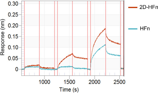
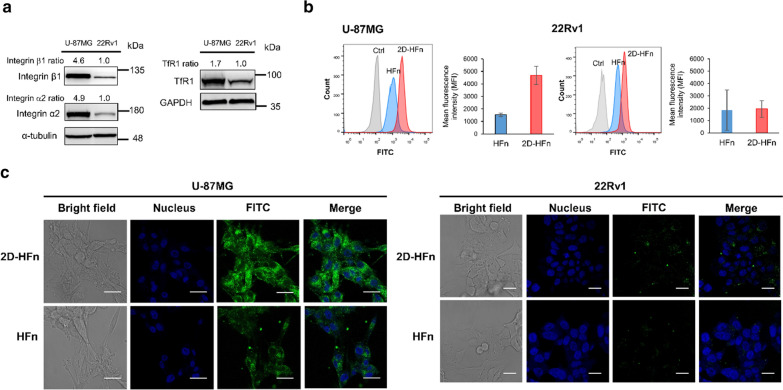
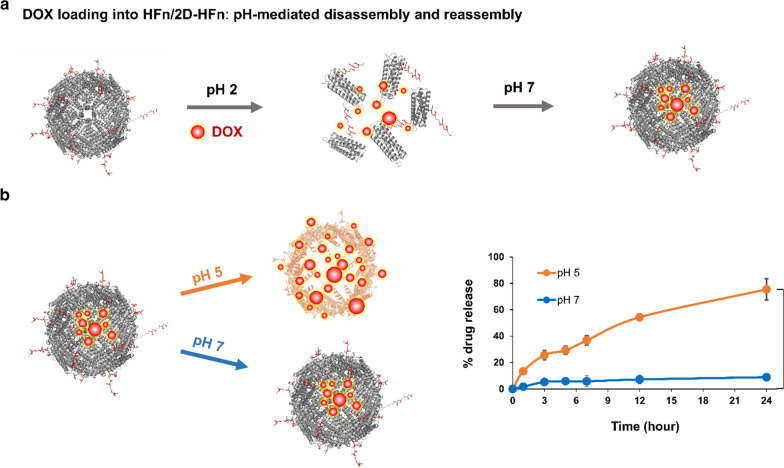
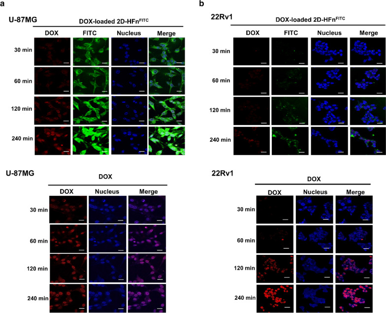
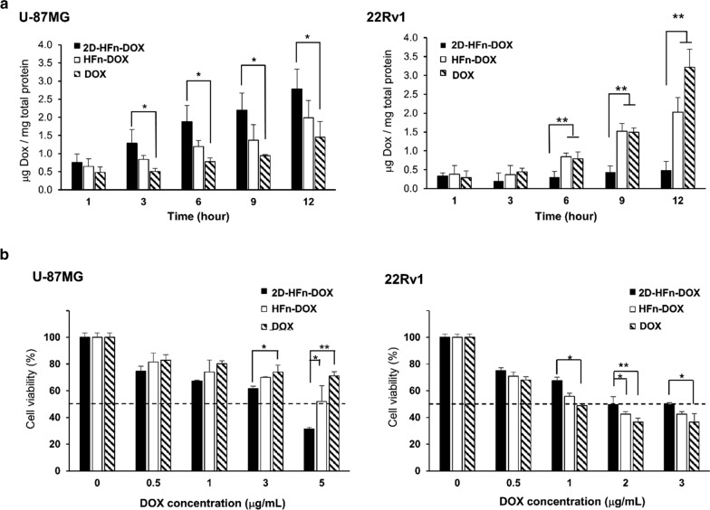
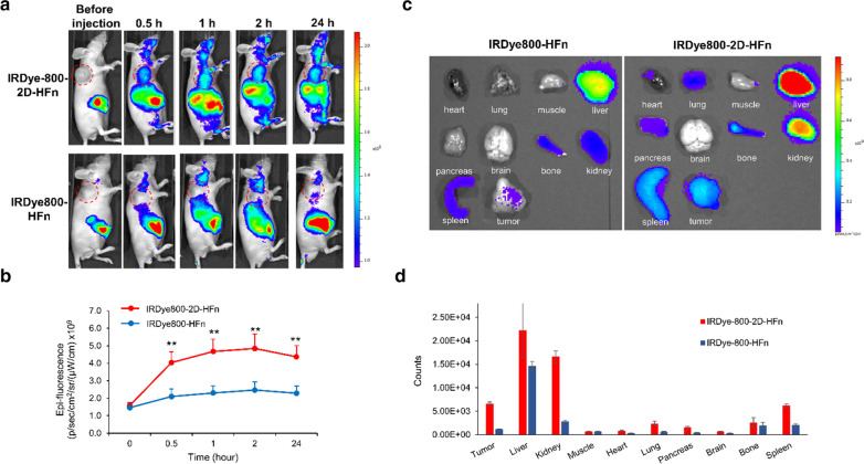
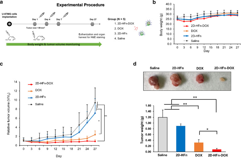
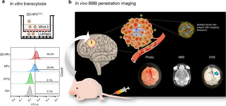
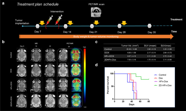
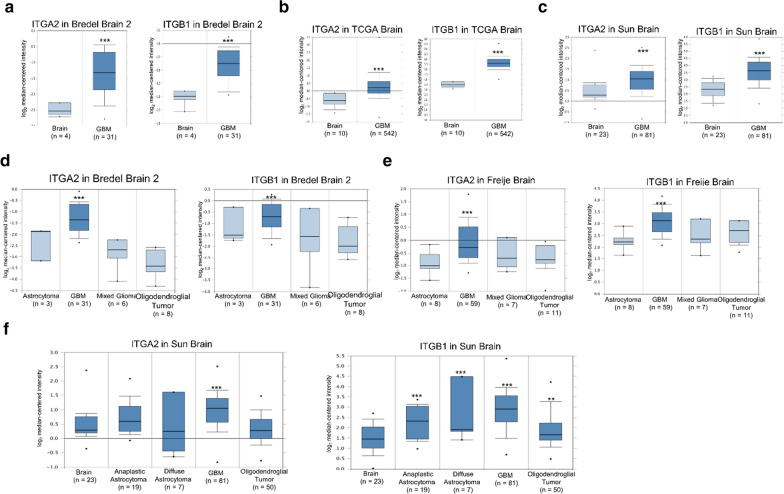
Results: In this study, a unique integrin α2β1-targeting H-ferritin (2D-HFn)-based drug delivery system was developed that highlights the feasibility of receptor-mediated transcytosis (RMT) for glioma tumor treatment. The integrin targeting α2β1 specificity was validated by biolayer interferometry in real time monitoring and followed by cell binding, chemo-drug encapsulation stability studies. Compared with naïve HFn, 2D-HFn dramatically elevated not only doxorubicin (DOX) drug loading capacity (up to 458 drug molecules/protein cage) but also tumor targeting capability after crossing BBB in an in vitro transcytosis assay (twofold) and an in vivo orthotopic glioma model. Most importantly, DOX-loaded 2D-HFn significantly suppressed subcutaneous and orthotopic U-87MG tumor progression; in particular, orthotopic glioma mice survived for more than 80 days.

Conclusions: We believe that this versatile nanoparticle has established a proof-of-concept platform to enable more accurate brain tumor targeting and precision treatment arrangements. Additionally, this unique RMT based ferritin drug delivery technique would accelerate the clinical development of an innovative drug delivery strategy for central nervous system diseases with limited side effects in translational medicine.

Keywords: Blood–brain barrier; Ferritin; Integrin α2β1; Receptor-mediated transcytosis (RMT); Transferrin receptor 1.


Conflict of interest statement

There are no conflicts to declare.

Figures

Fig. 1
Fig. 1
The validation of 2D-HFn and HFn binding to integrin α2β1 assayed with BLI. The integrin α2β1proteins were loaded to the sensor tip from a solution of 10 nM and then incubated with various concentrations of 2D-HFn and HFn in binding buffer with 2 mM Mg2+. The association and dissociation binding curves were monitored by eight-channel BLI system Octet RED96 and Kd values were calculated by Octet DataAnalysis software (9.0.0.6.)
Fig. 2
Fig. 2
2D-HFn has a strong ability to bind to cancer cells. a High expression of integrin α2β1 and TfR1 in U-87MG cells, but not in 22Rv1 cells by western blotting using antibodies against integrin α2, integrin β1 and TfR1. b FITC-labeled 2D-HFn had a stronger cell binding ability to U-87MG cells than FITC-labeled HFn. The mean fluorescence intensity (MFI) of FITC-labeled 2D-HFn on U-87MG cells was approximately 3.6-fold greater than that of FITC-labeled HFn (N = 3; mean ± SEM). c After incubation of FITC-labeled 2D-HFn with U-87MG cells, 2D-HFn bound to the cell surface and entered into the cells. In contrast, few 2D-HFn molecules bound to the cell surface of 22Rv1 cells. Scale bar, 10 μm
Fig. 3
Fig. 3
DOX was gradually released from DOX-loaded 2D-HFn in an acidic environment. a Encapsulation of DOX into 2D-HFn and HFn was performed by the pH-mediated disassembly and reassembly method. b A diagram showed the encapsulation of DOX into the ferritin complex and DOX was released at pH5 or pH7. DOX loaded inside DOX-2D-HFn at pH 7 was stable over 24 h, but 75% of the DOX was released from DOX-loaded 2D-HFn at pH 5 (**P < 0.01; paired t-test; N = 3; mean ± SEM)
Fig. 4
Fig. 4
2D-HFn specifically delivered DOX to cancer cells that highly express integrin α2β1. a DOX-loaded 2D-HFn nanoparticles or DOX was incubated with U-87MG cells for various times. The free form of DOX was inside the cell nuclei after 30 min of incubation. However, DOX delivered via the DOX-loaded 2D-HFn nanoparticle was inside the cell nucleus after two hours of incubation and became obvious after four hours of incubation. The red signal represents DOX; FITC represents 2D-HFn. b The same procedure was conducted with 22Rv1 cells. Free DOX entered the cell nuclei after two hours of incubation. However, few DOX and 2D-HFn signals were observed, even after four hours of DOX-loaded 2D-HFn incubation. Scale bar, 10 μm
Fig. 5
Fig. 5
2D-HFn enhanced the cytotoxic effects of DOX on cancer cells. a Cellular DOX uptake via DOX-loaded 2D-HFn, DOX-loaded HFn or free DOX in U-87MG and 22Rv1 cells (*P < 0.05, **P < 0.01; one-way ANOVA; N = 3; mean ± SEM). b Cytotoxicity of DOX-loaded 2D-HFn, DOX-loaded HFn or free DOX in U-87MG and 22Rv1 cells. Dashed lines indicated 50% cell viability. (*P < 0.05, **P < 0.01; one-way ANOVA; N = 3; mean ± SEM)
Fig. 6
Fig. 6
The tumor-targeting capability of 2D-HFn in U-87MG xenograft mouse models. a 2D-HFn had in vivo tumor-targeting capability in subcutaneous glioma mouse models. IRDye800-labeled 2D-HFn or IRDye800-labeled HFn was i.v. injected into U-87MG tumor-bearing mice, and the IRDye800 signal was acquired at various time points by an IVIS system. The dashed red circle represented the tumor location. b Quantitative values of the IRDye800 signal at different time points. Compared with HFn, the signal from 2D-HFn at the tumor site was approximately two-fold greater, and the tumor-targeting capability was enhanced (**P < 0.01; one-way ANOVA; N = 5; mean ± sd). c The biodistribution of 2D-HFn and HFn in U-87MG tumor-bearing mice. d The ROI signal analysis of the ex vivo organ imaging. The 2D-HFn signal in the tumors was stronger than the HFn signal in the tumors
Fig. 7
Fig. 7
DOX-loaded 2D-HFn remarkably reduced tumor growth. a The experimental procedure of the therapeutic treatment on U-87MG tumor-bearing mice. Briefly, mice were implanted with U-87MG cells via subcutaneous injection. When the tumor size reached approximately 80 mm3, the mice were randomly divided into four groups and i.v. injected with various treatments every three days for a total of three injections. The four groups included the saline (control), 2D-HFn, DOX and DOX-loaded 2D-HFn (2D-HFn-DOX) groups. The body weights and the tumor sizes were measured every two days. b There was no significant difference in body weight among all groups during the procedure. c The tumor volumes of each group during the treatment. The average tumor volume of the DOX-loaded 2D-HFn group was remarkably smaller than that of the other groups (*P < 0.05, **P < 0.01; one-way ANOVA; N = 5; mean ± sd). d After treatment, the mice were sacrificed, and the tumors from all groups were dissected and weighed. Compared with the other groups, the average tumor weight of the mice treated with DOX-loaded 2D-HFn was the smallest (*P < 0.05, **P < 0.01; one-way ANOVA; N = 5; mean ± sd)
Fig. 8
Fig. 8
In vitro crossing of the BBB & in vivo tumor-targeting imaging. a The in vitro transcytosis experiment was performed to assess the abilities of HFn and 2D-HFn to cross the BBB. Briefly, bEnd.3 mouse brain endothelial cells were cultured in the upper chamber of a Boyden chamber to mimic the BBB. U-87MG cells were cultured in a 6-well plate. FITC, FITC-HFn, or FITC-2D-HFn was added into the medium of the upper chamber. After four hours of incubation, the U-87MG cells were harvested, and the FITC signal was analyzed by flow cytometry. No signal was detected in the FITC group. Approximately 29.4% of the cells were FITC-positive in the FITC-HFn group, indicating that FITC-HFn crossed the bEnd.3 cell layers and bound to U-87MG cells. In comparison, 59.2% of the cells were FITC-positive in the FITC-2D-HFn group, approximately two-fold greater than that of the FITC-HFn group. b 2D-HFn had a strong tumor-targeting capability in an orthotopic tumor model. IRDye800-2D-HFn was iv injected into the orthotopic U-87MG tumor model mice, and the tumor volume and location of IRDye800-2D-HFn were analyzed by MRI and IVIS imaging, respectively
Fig. 9
Fig. 9
In vivo intracranial treatment, molecular imaging, and glioma mouse survival. a Treatment planning of the intracranial glioma tumor model. b-c In vivo PET/CT and MR imaging evaluation of glioma mice intravenously injected with saline (control), DOX, HFn-DOX or 2D-HFn-DOX and the quantitative analysis of anatomic tumor size and FDG uptake (N = 6). d Animal survival curves in the different treatment groups (Kaplan–Meier, P < 0.001)
Fig. 10
Fig. 10
Integrin α2 (ITGA2) and integrin β1 (ITGB1) expression were up-regulated in glioblastoma in clinical cancer samples in three datasets from the Oncomine online microarray database. The mRNA level of integrin α2 (ITGA2) and integrin β1 (ITGB1) in clinical brain tumor tissues and normal brain tissues were acquired and analyzed from the Oncomine online microarray database. A box-and-whisker plot that represents ITGA2 and ITGB1 expression. The horizontal top and bottom lines of each box represented the 75th and the 25th percentile, respectively. The band in the box is the median value. Horizontal lines above and below the box represented the 90th and the 10th percentile, respectively. The dots above the 90th percentile and below the 10th percentile represented the maximum and minimum values, respectively. ac From three brain datasets, ITGA2 and ITGB1 are up-regulated in GBM tissue specimens in comparison to normal tissues. df From three brain datasets, ITGA2 and ITGB1 are up-regulated in GBM tissue specimens in comparison to other subtypes of brain tumor tissue specimens and normal brain tissue specimens. Detailed information was listed in Additional file 1: Table S1. ** P < 0.01, *** P < 0.001

Similar articles

Cited by

References

    1. World Health Organization. Cancer. https://www.who.int/news-room/fact-sheets/detail/cancer.
    1. Park K. Controlled drug delivery systems: past forward and future back. J Control Release. 2014;190:3–8. doi: 10.1016/j.jconrel.2014.03.054. - DOI - PMC - PubMed
    1. Sun T, Zhang YS, Pang B, Hyun DC, Yang M, Xia Y. Engineered nanoparticles for drug delivery in cancer therapy. Angew Chem Int Ed Engl. 2014;53:12320–12364. - PubMed
    1. Dasari S, Tchounwou PB. Cisplatin in cancer therapy: molecular mechanisms of action. Eur J Pharmacol. 2014;740:364–378. doi: 10.1016/j.ejphar.2014.07.025. - DOI - PMC - PubMed
    1. Senapati S, Mahanta AK, Kumar S, Maiti P. Controlled drug delivery vehicles for cancer treatment and their performance. Signal Transduct Target Ther. 2018;3:7. doi: 10.1038/s41392-017-0004-3. - DOI - PMC - PubMed

MeSH terms

LinkOut - more resources