. 2021 Jun;28(6):3225-3234.
doi: 10.1016/j.sjbs.2021.02.061. Epub 2021 Feb 24.

Comparative anticancer activities of Ficus carica and Ficus salicifolia latex in MDA-MB-231 cells

Affiliations

Comparative anticancer activities of Ficus carica and Ficus salicifolia latex in MDA-MB-231 cells

Fatma Mousa AlGhalban et al. Saudi J Biol Sci. 2021 Jun.

Abstract

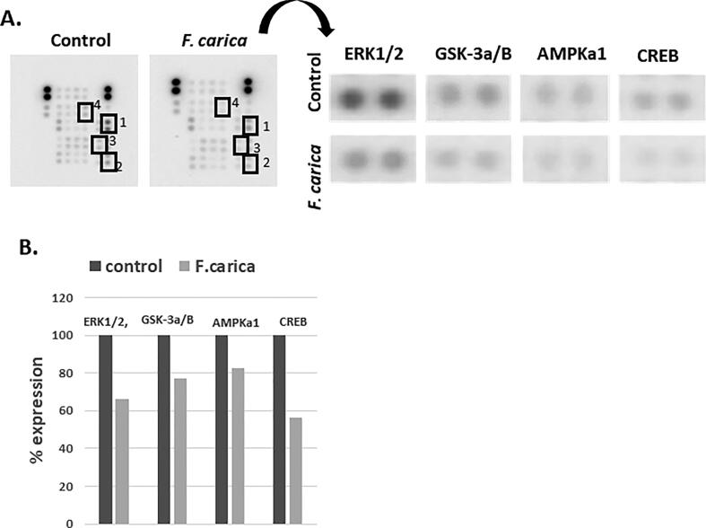
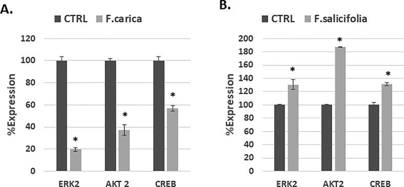
Ficus latex is rich in polyphenolic compounds and hence considered as an antioxidant and anti-proliferative. Many studies are available on Ficus carica (common fig) whereas Ficus salicifolia is less studied. F. salicifolia grows in a harsh dry environment, therefore its latex was selected in the current study along with the F. carica for their comparative anti-cancer potential and the involved molecular mechanism. Triple-negative breast cancer (TNBC) derived MDA-MB-231 cells were used in the study. MTT and morphological studies indicated that the latex of both plants has anti-proliferative effects. To know their anti-metastatic effects, a wound-healing assay was performed. Both species were able to maintain the wound size compared to the untreated cells indicating their anti-metastatic effects. Using a DNA damage assay kit, we found that both fig species have genotoxic and cytotoxic effects in MDA-MB-231 cells compared to the untreated control. To know the potential molecular mechanism involved, we used a human kinase array kit. We found that ERK2, CREB, and AKT2 were downregulated after treatment the MDA-Mb-231 cells with the latex of F. carica. We assumed that F. salicifolia will also affect the same pathways, however after confirmation through real-time (RT)-PCR, downregulations of the above mentioned pathways were confirmed in cells treated with F. carica latex, however, in cells treated with F. salicifolia the selected genes were upregulated at the transcriptional level. We conclude that latex of both species of ficus have anti-cancer effects in MDA-MB-231 cells, however differ in their level of toxicity and the mechanism of action at the molecular level.

Keywords: Anticancer; Ficus carica; Ficus salicifolia; Fig latex; MDA-MB-231.


Conflict of interest statement

The authors declare that they have no known competing financial interests or personal relationships that could have appeared to influence the work reported in this paper.

Figures

Fig. 1
Fig. 1
Flow chart representing the experimental design of the study.
Fig. 2
Fig. 2
The figure represents effects of (A) F. carica and (B) F. salicifolia latex on morphology of MDA-MB-231 cells after treatment with four different concentrations (0.1%, 0.25%, 0.5, 1%) and three time intervals (24 h, 48 h, 72 h). (C) Represents a zoom in for morphological changes caused by latex cell blebbing (c1, c5, c6), uneven cell shape (c2, c3), cytoplasmic vacuolation (c4).
Fig. 3
Fig. 3
The figure represents the cytotoxic effects of F. carica and F. salicifolia latex on viability in MDA-MB-231 cancer cells. The MDA-MB-231 cells were treated with latex of F. carica and F. salicifolia at four different concentrations (0.1, 0.25, 0.5, 1%) and three time points (24 h, 48 h, 72 h). A and B represent dose and time dependency for each treatment separately. C, D and E represent comparative cytotoxic effects of the two species at the different doses at 24 h, 48 h and 72 h respectively. The data are presented as Mean ± SD and statistical significance is accepted at p < 0.05. The symbols (a) represent control, (b) represents 0.1% dose, (c) represents 0.25% dose, (d) represents 0.5% dose and (e) represents 1.0% dose.
Fig. 4
Fig. 4
The figure represents effect of F. carica and F. salicifolia latex on on the MDA-MB-231 cancer cells migration. MDA-MB-231 cells treated with (0.01%, 0.025%, 0.05%) of F. carica and with (0.25%, 0.5%, 1%) of F. salicifolia for 24 h. The representative photographs of wound size at 0 h and 24 h for both F. carica (A) and F. salicifolia (C) are given. Figure B and D represents the quantified relative wound area after treatments with F. carica and F. salicifolia latex treatments.
Fig. 5
Fig. 5
The figure represents effects of F. carica and F. salicifolia latex on cytotoxicity and genotoxicity in MDA-MB-231 cells. The cells were treated with F. carica (0.1%) and F. salicifolia (1%) for 24 h. (A) represents cytotoxicity level in cells treated with F. carica and F. salicifolia latex and untreated cells using florescent microscopy and FTCI filter. (B) Represents quantified light intensity for FITC filter in all three groups. (C) Represents the genotoxicity of F. carica and F. salicifolia latex using florescent microscopy and DAPI staining. Red circles represent mitotic stages like metaphase and anaphase and white arrows represent stress morphologies like crescent shape, nucleus blebbing, uneven nucleus shape and chromatin condensation in addition to general nucleus shrinkage.
Fig. 6
Fig. 6
The figure shows effects of F. carica latex treatment on key proteins expression in MDA-MB-231 cells. (A) Represents difference in proteins expression and the proteins having difference in expression under ficus latex treatment compared to untreated controls are shown bound in squares. (B) Represents quantification of the proteins bound in square in A. The quantity was normalized with standard proteins expression.
Fig. 7
Fig. 7
Expression of ERK2, AKT2, CREB at transcriptional level after treatment with F. carica and F. salicifolia for 24 h RT-PCR was used to study the expression level of ERK2, AKT2 and CREB after they treated with (A) F. carica and (B) F. salicifolia.

References

    1. Badgujar S.B., Patel V.V., Bandivdekar A.H., Mahajan R.T. Traditional uses, phytochemistry and pharmacology of Ficus carica: a review. Pharm. Biol. 2014;52(11):1487–1503. - PubMed
    1. Bafor E.E., McKenna J., Rowan E.G., Edrada-Ebel R. Characterisation of the antiproliferative constituents and activity of Ficus exasperata (Vahl) on ovarian cancer cells–a preliminary investigation. Nat. Prod. Res. 2017;31(18):2164–2168. - PubMed
    1. Camero M., Marinaro M., Lovero A., Elia G., Losurdo M., Buonavoglia C., Tempesta M. In vitro antiviral activity of Ficus carica latex against caprine herpesvirus-1. Nat. Prod. Res. 2014;28(22):2031–2035. - PubMed
    1. Chavez K.J., Garimella S.V., Lipkowitz S. Triple negative breast cancer cell lines: one tool in the search for better treatment of triple negative breast cancer. Breast Dis. 2010;32(1–2):35. - PMC - PubMed
    1. Collignon J., Lousberg L., Schroeder H., Jerusalem G. Triple-negative breast cancer: treatment challenges and solutions. Breast Cancer: Targets Therapy. 2016;8:93. - PMC - PubMed

LinkOut - more resources