. 2021 May 26:15:657594.
doi: 10.3389/fncel.2021.657594. eCollection 2021.

Synaptic Remodeling in the Cone Pathway After Early Postnatal Horizontal Cell Ablation

Affiliations

Synaptic Remodeling in the Cone Pathway After Early Postnatal Horizontal Cell Ablation

Lena Nemitz et al. Front Cell Neurosci. .

Abstract

The first synapse of the visual pathway is formed by photoreceptors, horizontal cells and bipolar cells. While ON bipolar cells invaginate into the photoreceptor terminal and form synaptic triads together with invaginating horizontal cell processes, OFF bipolar cells make flat contacts at the base of the terminal. When horizontal cells are ablated during retina development, no invaginating synapses are formed in rod photoreceptors. However, how cone photoreceptors and their synaptic connections with bipolar cells react to this insult, is unclear so far. To answer this question, we specifically ablated horizontal cells from the developing mouse retina. Following ablation around postnatal day 4 (P4)/P5, cones initially exhibited a normal morphology and formed flat contacts with OFF bipolar cells, but only few invaginating contacts with ON bipolar cells. From P15 on, synaptic remodeling became obvious with clustering of cone terminals and mislocalized cone somata in the OPL. Adult cones (P56) finally displayed highly branched axons with numerous terminals which contained ribbons and vesicular glutamate transporters. Furthermore, type 3a, 3b, and 4 OFF bipolar cell dendrites sprouted into the outer nuclear layer and even expressed glutamate receptors at the base of newly formed cone terminals. These results indicate that cones may be able to form new synapses with OFF bipolar cells in adult mice. In contrast, cone terminals lost their invaginating contacts with ON bipolar cells, highlighting the importance of horizontal cells for synapse maintenance. Taken together, our data demonstrate that early postnatal horizontal cell ablation leads to differential remodeling in the cone pathway: whereas synapses between cones and ON bipolar cells were lost, new putative synapses were established between cones and OFF bipolar cells. These results suggest that synapse formation and maintenance are regulated very differently between flat and invaginating contacts at cone terminals.

Keywords: bipolar cells; cones; horizontal cells; photoreceptors; retina; ribbon synapse; synaptic remodeling; vision.


Conflict of interest statement

The authors declare that the research was conducted in the absence of any commercial or financial relationships that could be construed as a potential conflict of interest.

Figures

FIGURE 1
FIGURE 1
Horizontal cell ablation. (A,B) Retinal cryosections of Cx57+/+ and Cx57+/DTR mice (P56) were labeled with an antibody specific for the horizontal cell marker calbindin. In Cx57+/DTR mice, horizontal cells were completely lost, while calbindin-positive amacrine and ganglion cells were unaffected (B). Scale bar, 50 μm.
FIGURE 2
FIGURE 2
Mislocalized cone somata following early postnatal horizontal cell ablation. (A–F) Retinae of Cx57+/+ and Cx57+/DTR mice of different ages (P8, P15, and P21) were stained for cone arrestin, a marker for cone photoreceptors. In wild-type mice, cones displayed the classical morphology including outer segment, inner segment, cell body, axon and synaptic terminal (A–C). Cone somata were scattered across the entire ONL at P8 (A) and positioned in the distal part of the ONL from P15 to P21 (B,C). In Cx57+/DTR mice, the cone morphology was initially comparable to that in wild-type mice (A,D). From P15 onward, cone somata were partly mislocalized (arrowheads) (E,F) and cone terminals were irregularly distributed in the OPL (E,F). Scale bar, 50 μm.
FIGURE 3
FIGURE 3
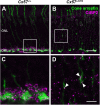
Cone neurite sprouting in adult horizontal cell-ablated mice. (A–D) Vertical sections of Cx57+/+ and Cx57+/DTR mice (P56) were labeled with antibodies against cone arrestin. While cones in wild-type mice had one axon with a single terminal (A,B), cones in horizontal cell-ablated mice had branched axons with numerous terminals (open arrowheads) (C,D). In some cases, several axons emerged from one soma (white arrowheads) (C,D). (E) Quantification of cone somata in vertical sections of wild-type (n = 3) and horizontal cell-ablated retinae (n = 3) (P56). p = 0.9736, t-test. Values are presented as mean ± SD. Scale bars, 25 μm (C), 10 μm (D).
FIGURE 4
FIGURE 4
Clustering and reduced gap junctional coupling of cone terminals in horizontal cell-ablated mice. (A–L) Immunolabeling of retinal whole mounts from Cx57+/+ and Cx57+/DTR mice with antibodies against cone arrestin. In the OPL of wild-type mice, cone pedicles formed a regular mosaic (A,B,E,F,I,J), whereas in horizontal cell-ablated mice, cone pedicles were unevenly spaced and several mislocalized cone somata were apparent (arrowheads) (C,D,G,H,K,L). (M–P) Vertical sections of Cx57+/+ and Cx57+/DTR retinae were stained for cone arrestin (green) and Cx36 (magenta). Cx36 expression in the OPL of horizontal cell-ablated mice was strongly reduced compared to wild-type mice. Scale bars, 25 μm (K), 10 μm (L,P).
FIGURE 5
FIGURE 5
Ribbons in new cone terminals. (A–D) Double staining of vertical sections from Cx57+/+ and Cx57+/DTR mice (P56) for cone arrestin, a cone marker (green), and CtBP2 (magenta), a synaptic ribbon marker. In the outer retina of wild-type mice, ribbons were horseshoe shaped and confined to the OPL (A,C). By contrast, ribbons in horizontal cell-ablated mice were smaller and distributed over the entire ONL (B,D). In addition, CtBP2-postive structures were frequently found in ectopic cone terminals (arrowheads) (D). Scale bars, 20 μm (B), 5 μm (D).
FIGURE 6
FIGURE 6
New cone terminals express vesicular glutamate transporters. (A–L) Retinal cryosections of Cx57+/+ and Cx57+/DTR mice (P56) were stained for the vesicular glutamate transporter VGluT1 (green) and cone arrestin (magenta). In wild-type and horizontal cell-ablated mice, cone pedicles in the OPL were VGluT1-positive (A–I). Moreover, VGluT1 immunoreactivity was present within ectopic cone terminals in the ONL of Cx57+/DTR mice (arrowheads) (J,K,L). Scale bars, 25 μm (C,I), 10 μm (F,L).
FIGURE 7
FIGURE 7
Loss of ON bipolar cell invaginations in cone terminals. (A–L) Electron microscopic analysis of individual cone terminals from Cx57+/+ and Cx57+/DTR mice at different ages (P11, P15, and P56). Cone terminals (yellow) in wild-type mice contained triads composed of horizontal cell dendrites (blue) and ON bipolar cell dendrites (red) as early as P11 (A,E,I). In horizontal cell-ablated mice, some cone terminals with horizontal cell invaginations were found at P11 (B). However, triads were rarely observed (C) and most cone pedicles contained no invaginations (D). At P15 and P56, cone terminals were generally lacking any invaginations in Cx57+/DTR mice (F–H,J–L). Ribbons were often shorter and free-floating (arrowheads) (B–D,F–H,J–L). Scale bar, 1 μm.
FIGURE 8
FIGURE 8
Absence of functional synapses between cones and ON bipolar cells in adult horizontal cell-ablated mice. (A–H) Triple labeling of retinae from Cx57+/+ and Cx57+/DTR mice (P56) for GPR179 (a component of the mGluR6 macromolecular complex, yellow), cone arrestin (a marker for cones, blue) and SCGN (a marker for a subset of ON and OFF bipolar cells, magenta). In wild-type mice, GPR179-immunoreactive puncta were observed at contact points between cone pedicles and bipolar cell dendrites (arrowheads) (A–D). In contrast, GPR179 labeling was completely absent in Cx57+/DTR mice (E–H), suggesting that adult horizontal cell-ablated mice lack functional synapses between cones and ON bipolar cells. Scale bar, 10 μm.
FIGURE 9
FIGURE 9
OFF bipolar cells sprouted into the ONL. (A–F) Retinal sections from Cx57+/+ and Cx57+/DTR retinae (P56) were stained for HCN4, PKARIIβ and calsenilin, markers for type 3a, 3b, and 4 OFF bipolar cells, respectively. While the dendrites of type 3a, 3b, and 4 OFF bipolar cells terminated in the OPL in wild-type mice (A–C), all three types showed an extensive outgrowth of dendrites into the ONL in horizontal cell-ablated mice (arrowheads) (D–F). Scale bar, 50 μm.
FIGURE 10
FIGURE 10
Cones established new synapses with OFF bipolar cells. (A–H’) Double labeling of vertical sections from Cx57+/+ and Cx57+/DTR retinae for the kainate receptor subunit GluK1 and the cone marker cone arrestin. In wild-type and horizontal cell-ablated mice, GluK1 immunoreactivity was found below the base of the cone terminals in the OPL from P8 to P56 (A–H). In Cx57+/DTR retinae, at P56, outgrowing GluK1-positive dendrites made contacts with the newly formed cone terminals in the ONL (arrowheads) (H’). (I–N) Double staining of retinal whole mounts from Cx57+/+ (K) and Cx57+/DTR mice (P56) (L–N) for GluK1 and cone arrestin. At the level of the OPL, cone terminals were associated with GluK1 staining in both genotypes (I,J). Ectopic cone terminals in the ONL of horizontal cell-ablated mice were directly connected to GluK1-positive dendrites (arrowheads) (K–N). Scale bars, 20 μm (H,K), 10 μm (H’,N).
FIGURE 11
FIGURE 11
Schematic illustration of rod and cone synaptogenesis in wild-type and horizontal cell-ablated mice. In wild-type mice, the development of rod and cone synapses begins with the formation of a contact between the photoreceptor terminal (yellow) and a single horizontal cell process (blue). Subsequently, a second horizontal cell process is recruited and both horizontal cell processes invaginate into the presynaptic terminal. In the last step, one or two ON bipolar cell dendrites (red) invaginate into the photoreceptor terminal and OFF bipolar cells (violet) form flat contacts at the base of the photoreceptor terminal. In the absence of horizontal cells, ON bipolar cells do not invaginate into rod and cone terminals and rod bipolar cells sprout into the ONL. Compared to wild-type mice, horizontal cell-ablated mice display fewer and shorter presynaptic ribbons, which are often not anchored to the photoreceptor membrane. At later stages, cones form new terminals with synaptic ribbons which are contacted by sprouting OFF bipolar cell dendrites. (Note that synapses between rod terminals and OFF bipolar cells have not been investigated for Cx57+/+ and Cx57+/DTR mice.)

Similar articles

Cited by

References

    1. Behrens C., Schubert T., Haverkamp S., Euler T., Berens P. (2016). Connectivity map of bipolar cells and photoreceptors in the mouse retina. eLife 5:e20041. 10.7554/eLife.20041 - DOI - PMC - PubMed
    1. Blanks J. C., Adinolfi A. M., Lolley R. N. (1974). Synaptogenesis in the photoreceptor terminal of the mouse retina. J. Comp. Neurol. 156 81–93. 10.1002/cne.901560107 - DOI - PubMed
    1. Chong N. H., Alexander R. A., Barnett K. C., Bird A. C., Luthert P. J. (1999). An immunohistochemical study of an autosomal dominant Feline Rod/Cone dysplasia (Rdy Cats). Exp. Eye Res. 68 51–57. 10.1006/exer.1998.0580 - DOI - PubMed
    1. Dick O., tom Dieck S., Altrock W. D., Ammermüller J., Weiler R., Garner C. C., et al. (2003). The presynaptic active zone protein bassoon is essential for photoreceptor ribbon synapse formation in the retina. Neuron 37 775–786. 10.1016/S0896-6273(03)00086-2 - DOI - PubMed
    1. Fariss R. N., Li Z.-Y., Milam A. H. (2000). Abnormalities in rod photoreceptors, amacrine cells, and horizontal cells in human retinas with retinitis pigmentosa. Am. J. Ophthalmol. 129 215–223. 10.1016/S0002-9394(99)00401-8 - DOI - PubMed