. 2021 May 28:15:682383.
doi: 10.3389/fnana.2021.682383. eCollection 2021.

Maturation-Dependent Differences in the Re-innervation of the Denervated Dentate Gyrus by Sprouting Associational and Commissural Mossy Cell Axons in Organotypic Tissue Cultures of Entorhinal Cortex and Hippocampus

Affiliations

Maturation-Dependent Differences in the Re-innervation of the Denervated Dentate Gyrus by Sprouting Associational and Commissural Mossy Cell Axons in Organotypic Tissue Cultures of Entorhinal Cortex and Hippocampus

Mandy H Paul et al. Front Neuroanat. .

Abstract

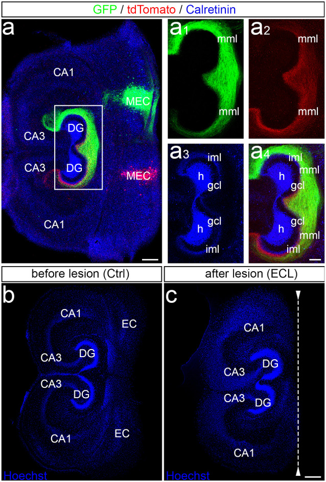
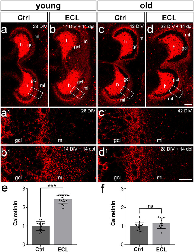
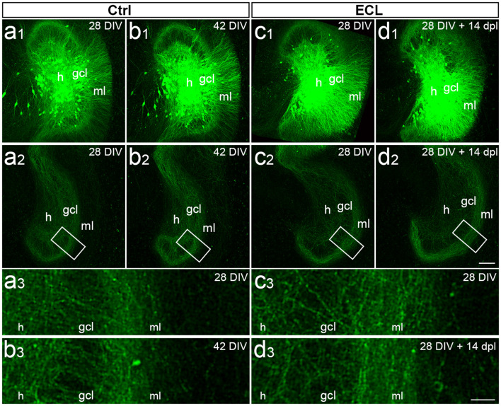
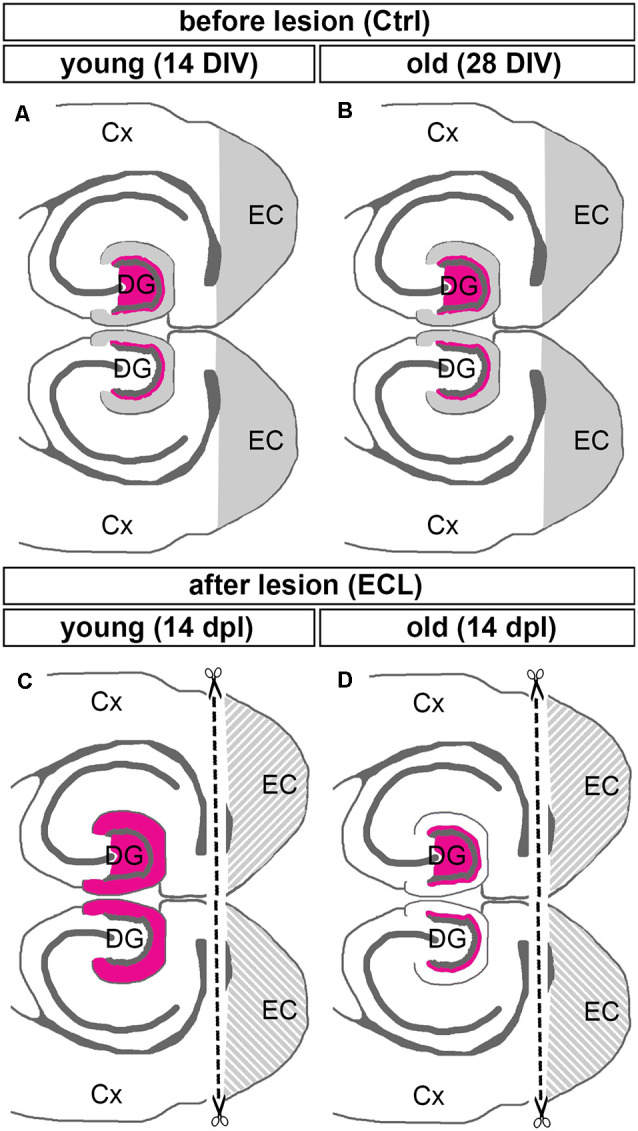
Sprouting of surviving axons is one of the major reorganization mechanisms of the injured brain contributing to a partial restoration of function. Of note, sprouting is maturation as well as age-dependent and strong in juvenile brains, moderate in adult and weak in aged brains. We have established a model system of complex organotypic tissue cultures to study sprouting in the dentate gyrus following entorhinal denervation. Entorhinal denervation performed after 2 weeks postnatally resulted in a robust, rapid, and very extensive sprouting response of commissural/associational fibers, which could be visualized using calretinin as an axonal marker. In the present study, we analyzed the effect of maturation on this form of sprouting and compared cultures denervated at 2 weeks postnatally with cultures denervated at 4 weeks postnatally. Calretinin immunofluorescence labeling as well as time-lapse imaging of virally-labeled (AAV2-hSyn1-GFP) commissural axons was employed to study the sprouting response in aged cultures. Compared to the young cultures commissural/associational sprouting was attenuated and showed a pattern similar to the one following entorhinal denervation in adult animals in vivo. We conclude that a maturation-dependent attenuation of sprouting occurs also in vitro, which now offers the chance to study, understand and influence maturation-dependent differences in brain repair in these culture preparations.

Keywords: calretinin; dentate gyrus; layer-specificity; organotypic culture; perforant path transection.


Conflict of interest statement

The authors declare that the research was conducted in the absence of any commercial or financial relationships that could be construed as a potential conflict of interest.

Figures

Figure 1
Figure 1
Entorhinal projection and entorhinal lesion in vitro. (A) Viral tracing was employed to visualize the entorhino-hippocampal projection in double cultures. One medial entorhinal cortex (MEC) was injected with an adeno-associated virus transducing a green construct (AAV2-hSyn-GFP) and the other MEC was injected with a virus transducing a red construct (AAV2-hSyn-tdTomato). In addition, calretinin-immunofluorescence labeling was used to visualize the mossy cells and their axons (blue; overexposed channel to visualize the axons in the inner molecular layer). CA3, CA1, hippocampal subfields CA3, CA1; DG, dentate gyrus; h, hilus; gcl, granule cell layer; iml, inner molecular layer; mml, middle molecular layer. Scale bar: 200 μm. (A1–A4) Higher magnification of the dentate gyrus (DG) showing the termination pattern of green MEC axons (A1), red MEC axons (A2), calretinin-immunolabeled axons (A3), and of all three projections (A4). Scale bar: 100 μm (A1–A4). (B,C) Entorhinal denervation was performed under visual control. A section (30 μm) of an old (DIV 28) double entorhino-hippocampal tissue culture is illustrated before (B) and after (C) lesion. Sections were fixed and nuclear staining with Hoechst was used to show that the entorhinal lesion leaves the DG and hippocampus proper intact. EC, entorhinal cortex. Scale bars: 250 μm (B,C).
Figure 2
Figure 2
Commissural/associational sprouting is attenuated in cultures lesioned at 28 days in vitro. (A–D) Double cultures of wild type mouse hippocampus immunolabeled for calretinin (red). (A) Overview of a non-denervated young double culture at DIV 14 (control, Ctrl). Calretinin-positive mossy cells were found in the hilus (h) of the culture and mossy cell axons were present in the inner part of the molecular layer (ml). (B) Fourteen days post-lesion (dpl) mossy cell axons were found throughout the ml. (A1,B1) Higher magnification of the boxed areas in (A,B) reveals a robust ingrowth of mossy cell axons into the outer part of the ml (A1 compared to B1) after entorhinal cortex lesion (ECL). (C) Overview of a non-denervated old double culture at DIV 28 (Ctrl). Calretinin-positive mossy cells were found in the hilus (h) of the culture, while mossy cell axons were found in the inner part of the molecular layer (ml). (D) Fourteen days post-lesion (dpl) mossy cell axons in old cultures stayed within the inner zone of the molecular layer. (C1,D1) Higher magnification of the boxed areas in (C,D) reveals only a very limited sprouting response (C1 compared to D1) after ECL. Compared to young cultures (B1 compared to D1), the sprouting response is greatly attenuated. (E) Quantitative analysis of the axonal sprouting response into the ml in young cultures. Control (Ctrl) was set to 1. A highly significant sprouting response was seen in denervated young cultures compared to non-denervated control cultures (Ctrl: 1.00 ± 0.24, ECL: 2.45 ± 0.22). (F) Quantitative analysis of the axonal sprouting response into the ml in old cultures. Control (Ctrl) was set to 1. No significant (ns) sprouting response was seen in denervated old cultures compared to non-denervated control cultures (Ctrl: 1.00 ± 0.21, ECL: 1.16 ± 0.28). Mann–Whitney test; mean ± SD; ***p < 0.001; n = 14–18 hippocampi per group in young cultures (data of young cultures from Del Turco et al., 2019) and n = 12–14 hippocampi per group in old cultures; gcl, granule cell layer. Scale bars: 100 μm (A–D); 25 μm (A1–D1).
Figure 3
Figure 3
Viral tracing and time-lapse imaging confirm attenuated sprouting response of older cultures. (A–D) Imaging of old (DIV 28) entorhino-hippocampal double cultures without (Ctrl; A,B) and with entorhinal cortex lesion (ECL; C,D). At DIV 4 both sets of cultures received an injection of AAV2-hSyn1-GFP into one hilus (h). On 28 days in vitro (DIV) a subset of cultures served as non-lesioned time-matched controls (A,B), whereas a second subset of cultures received an ECL (C,D). (A1,B1) Images of the same virus injected control culture at DIV 28 (A1) and DIV 42 (B1). Commissural axons in the contralateral dentate gyrus are illustrated at DIV 28 (A2) and DIV 42 (B2) respectively. The boxed areas are shown at higher magnification (A3,B3), illustrating minimal changes occurring during the 14 days observation period. (C1,D1) Images of the same virus injected culture at DIV 28 (C1) and 14 days post-lesion (dpl) (D1). Commissural axons in the contralateral dentate gyrus are shown at DIV 28 (C2) and DIV 28 + 14 dpl (D2). The boxed areas are shown at higher magnification (C3,D3) illustrating no major differences between DIV 28, DIV 42, and 14 dpl. gcl, granule cell layer. Scale bar: 100 μm (A1–D1 and A2–D2); 25 μm (A3–D3).
Figure 4
Figure 4
Summary diagram illustrating maturation-dependent differences in the sprouting of commissural/associational mossy cell axons after entorhinal lesion. (A,B) Before lesion (control, Ctrl) DIV 14 (young) and DIV 28 (old) double entorhino-hippocampal tissue cultures showed an organotypic and layer-specific entorhinal innervation of the outer molecular layer of the dentate gyrus (DG, gray). Commissural/associational axons arising from hilar mossy cells terminate in the inner molecular layer of the dentate gyrus (magenta; only the mossy cell projection of the upper DG is shown). (C,D) Fourteen days post-lesion (dpl) a robust sprouting response is seen in young cultures (C). In these cultures, commissural/associational axons grow into the denervated zone up to the hippocampal fissure. This corresponds to the juvenile sprouting response reported for commissural/associational axons in vivo. (D) In contrast, in older cultures commissural/associational axons stayed largely in their home territory and did not invade the denervated zone. This corresponds to the adult pattern of sprouting of commissural/associational axons seen in vivo. Note, only the mossy cell projection of the upper DG is shown in (C,D). Cx, cortex; EC, entorhinal cortex.

Similar articles

Cited by

References

    1. Askvig J. M., Watt J. A. (2019). Absence of axonal sprouting following unilateral lesion in 125-day-old rat supraoptic nucleus may be due to age-dependent decrease in protein levels of ciliary neurotrophic factor receptor alpha. J. Comp. Neurol. 527, 2291–2301. 10.1002/cne.24675 - DOI - PMC - PubMed
    1. Caroni P. (1997). Intrinsic neuronal determinants that promote axonal sprouting and elongation. Bioessays 19, 767–775. 10.1002/bies.950190906 - DOI - PubMed
    1. Cotman C. W., Nadler J. V. (1978). “Reactive synaptogenesis in the hippocampus,” in Neuronal Plasticity, ed Cotman C. W. (New York, NY: Raven Press; ), 227–271.
    1. Dailey M. E., Smith S. J. (1996). The dynamics of dendritic structures in developing hippocampal slices. J. Neurosci. 16, 2983–2994. 10.1523/JNEUROSCI.16-09-02983.1996 - DOI - PMC - PubMed
    1. Del Turco D., Deller T. (2007). Organotypic entorhino-hippocampal slice cultures–a tool to study the molecular and cellular regulation of axonal regeneration and collateral sprouting in vitro. Methods Mol. Biol. 399, 55–66. 10.1007/978-1-59745-504-6_5 - DOI - PubMed

LinkOut - more resources