

Artificial Intelligence Can Effectively Predict Early Hematoma Expansion of Intracerebral Hemorrhage Analyzing Noncontrast Computed Tomography Image
- PMID: 34122038
- PMCID: PMC8188896
- DOI: 10.3389/fnagi.2021.632138
Artificial Intelligence Can Effectively Predict Early Hematoma Expansion of Intracerebral Hemorrhage Analyzing Noncontrast Computed Tomography Image
Abstract
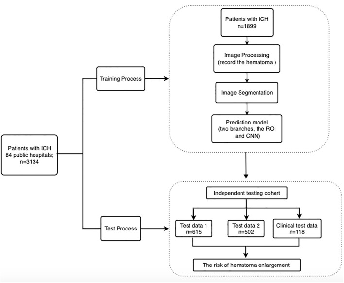
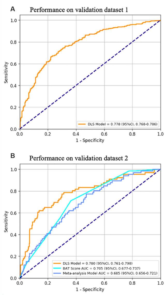
This study aims to develop and validate an artificial intelligence model based on deep learning to predict early hematoma enlargement (HE) in patients with intracerebral hemorrhage. A total of 1,899 noncontrast computed tomography (NCCT) images of cerebral hemorrhage patients were retrospectively analyzed to establish a predicting model and 1,117 to validate the model. And a total of 118 patients with intracerebral hemorrhage were selected based on inclusion and exclusion criteria so as to validate the value of the model for clinical prediction. The baseline noncontrast computed tomography images within 6 h of intracerebral hemorrhage onset and the second noncontrast computed tomography performed at 24 ± 3 h from the onset were used to evaluate the prediction of intracerebral hemorrhage growth. In validation dataset 1, the AUC was 0.778 (95% CI, 0.768-0.786), the sensitivity was 0.818 (95% CI, 0.790-0.843), and the specificity was 0.601 (95% CI, 0.565-0.632). In validation dataset 2, the AUC was 0.780 (95% CI, 0.761-0.798), the sensitivity was 0.732 (95% CI, 0.682-0.788), and the specificity was 0.709 (95% CI, 0.658-0.759). The sensitivity of intracerebral hemorrhage hematoma expansion as predicted by an artificial intelligence imaging system was 89.3%, with a specificity of 77.8%, a positive predictive value of 55.6%, a negative predictive value of 95.9%, and a Yoden index of 0.671, which were much higher than those based on the manually labeled noncontrast computed tomography signs. Compared with the existing prediction methods through computed tomographic angiography (CTA) image features and noncontrast computed tomography image features analysis, the artificial intelligence model has higher specificity and sensitivity in the prediction of early hematoma enlargement in patients with intracerebral hemorrhage.
Keywords: artificial intelligence; convolutional neural network; hematoma expansion; intracerebral hematoma; predict.
Copyright © 2021 Teng, Ren, Zhang, Wu, Guo and Ren.
Conflict of interest statement
Authors PZ and ZW were employed by company BioMind Technology. The remaining authors declare that the research was conducted in the absence of any commercial or financial relationships that could be construed as a potential conflict of interest.
Figures
References
-
- Boulouis G., Morotti A., Brouwers H. B., Charidimou A., Jessel M. J., Auriel E., et al. . (2016). Association between hypodensities detected by computed tomography and hematoma expansion in patients with intracerebral hemorrhage. JAMA Neurol. 73, 961–968. 10.1001/jamaneurol.2016.1218 - DOI - PMC - PubMed
-
- Brouwers H. B., Battey T. W., Musial H. H., Ciura V. A., Falcone G. J., Ayres A. M., et al. . (2015). Rate of contrast extravasation on computed tomographic angiography predicts hematoma expansion and mortality in primary intracerebral hemorrhage. Stroke 46, 2498–2503. 10.1161/STROKEAHA.115.009659 - DOI - PMC - PubMed
LinkOut - more resources
Full Text Sources