. 2021 May 28:12:667282.
doi: 10.3389/fphar.2021.667282. eCollection 2021.

Selective Wnt/β-Catenin Pathway Activation Concomitant With Sustained Overexpression of miR-21 is Responsible for Aristolochic Acid-Induced AKI-to-CKD Transition

Affiliations

Selective Wnt/β-Catenin Pathway Activation Concomitant With Sustained Overexpression of miR-21 is Responsible for Aristolochic Acid-Induced AKI-to-CKD Transition

Qing Kuang et al. Front Pharmacol. .

Abstract

Acute kidney injury (AKI) is increasingly recognized as a cumulative risk factor for chronic kidney disease (CKD) progression. However, the underlying mechanisms remain unclear. Using an aristolochic acid (AA)-induced mouse model of AKI-to-CKD transition, we found that the development of tubulointerstitial fibrosis following AKI was accompanied with a strong activation of miR-21 and canonical Wnt signaling, whereas inhibition of miR-21 or selective silencing of Wnt ligands partially attenuated AKI-to-CKD transition. To explore the interaction between miR-21 and Wnt/β-catenin signaling, we examined the effects of genetic absence or pharmacologic inhibition of miR-21 on Wnt/β-catenin pathway expression. In miR-21-/- mice and in wild-type mice treated with anti-miR21 oligos, Wnt1 and Wnt4 canonical signaling in the renal tissue was significantly reduced, with partial reversal of renal interstitial fibrosis. Although the renal abundance of miR-21 remained unchanged after inhibition or activation of Wnt/β-catenin signaling, early intervention with ICG-001, a β-catenin inhibitor, significantly attenuated renal interstitial fibrosis. Moreover, early (within 24 h), but not late β-catenin inhibition after AA administration attenuated AA-induced apoptosis and inflammation. In conclusion, inhibition of miR-21 or β-catenin signaling may be an effective approach to prevent AKI-to-CKD progression.

Keywords: AKI-CKD transition; Wnt; aristolochic acid; miR-21; β-catenin.


Conflict of interest statement

The authors declare that the research was conducted in the absence of any commercial or financial relationships that could be construed as a potential conflict of interest.

Figures
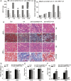
FIGURE 1
FIGURE 1
Inhibition of miR⁃21 partially reverses renal tubulointerstitial fibrosis induced by aristolochic acid. (A,B) RT-qPCR analysis of miR-21 abundance and serum levels of creatinine. Mice were sacrificed on days 1, 3, 7, 14, or 28 after aristolochic acid (AA) administration (10 mg/kg, i.p.). Locked nucleic acid- (LNA-) modified anti-miR21 oligonucleotides or anti-scramble was administered via the tail vein (10 mg/kg, 5 mg/ml) within 60 min prior to AA delivery. Additional injections (the same dose) were given on the 5th and 10th day after AA-I dosing. *p < 0.05 and **p < 0.01 vs. anti-scramble + AA groups, n = 6. (C) H&E and Masson’s trichrome staining of the corticomedullary junction (magnification, 400×, bar = 200 μm) on days 14 and 28 after AA delivery. (D,E) Histopathological assessment of H&E and Masson’s trichrome staining. Ten high-power fields for each mouse were captured, and then the average number in the high-power fields was calculated for quantification. (F) The miR-21 abundance. *p < 0.05 and **p < 0.01 vs. control groups (Ctrl). # p < 0.05 and ## p < 0.01 vs. anti-scramble + AA groups, n = 6.
FIGURE 2
FIGURE 2
Sustained Wnt/β-catenin pathway activation secondary to AA induces AKI. (A) Western blot of representative Wnt ligands (Wnt1, Wnt2b, Wnt3, Wnt4, Wnt7a) and β-catenin in mouse kidney tissues. Mice were sacrificed on days 1, 3, 7, 14, or 28 after AA administration. (B–G) Quantitative western blot data. (H) Immunohistochemical staining of Wnt1 and Wnt4 in the kidneys (magnification, 400×, bar = 200 μm). (I,J) The number of Wnt1/Wnt4 positive cells in the mouse kidneys. Each mouse was calculated for quantification of 10 high-power fields. *p < 0.05 and **p < 0.01 vs. control groups (Ctrl), n = 6.
FIGURE 3
FIGURE 3
Inhibition of miR-21 selectively suppresses Wnt/β-catenin signaling in vivo and vitro. (A–G) Western blots and quantitative data of renal expression of Wnt1, Wnt2b, Wnt3, Wnt4, Wnt7a, and β-catenin on days 14 and 28 after AA delivery. LNA-modified anti-miR21 oligonucleotides or anti-scramble was administered to the mice via the tail vein (10 mg/kg, 5 mg/ml) within 60 min prior to AA delivery. Additional injections (the same dose) were given on the 5th and 10th day after AA-I dosing. (H–J) Immunohistochemical staining of Wnt1 and Wnt4 in the kidneys (magnification, 400×, bar = 200 μm) and the number of Wnt1/Wnt4 positive cells in the mouse kidneys. *p < 0.05, **p < 0.01 vs. controls. # p < 0.05, ## p < 0.01 vs. anti-scrambled control groups, n = 6. (K–M) Changes in protein abundances of collagen IV, α-SMA, vimentin, Wnt1, Wnt4, and β-catenin and miR-21 abundance in HK-2 cells. HK-2 cells were pretreated with 50 nM anti-miR21 or 50 nM anti-scramble 6 h prior to 48 h-AA treatment (2 μg/ml). *p < 0.05 and **p < 0.01 vs. anti-scramble controls, n = 3.
FIGURE 4
FIGURE 4
Absence of miR-21 hinders AKI-to-CKD progression by selectively downregulating the Wnt/β-catenin pathway. (A) Renal miR-21 abundance. The wild-type mice and miR-21 knockout mice were sacrificed on days 7 and 14 after AA administration. (B) Serum creatinine levels of mice. (C) Kidney injury scores of mice on days 7 and 14 post AA delivery. (D) Collagen abundance was analyzed using Masson’s trichrome staining. (E) Renal histologic patterns of wild-type mice and mir-21 KO mice (magnification, 200×, bar = 200 μm). (F–L) Relative protein expression of Wnt1, Wnt4, β-catenin, α-SMA, Wnt2b, and collagen I in kidneys. *p < 0.05, **p < 0.01 vs. controls. # p < 0.05, ## p < 0.01 vs. KO + AA, n = 3.
FIGURE 5
FIGURE 5
The timing of β-catenin inhibition determines the reversibility of AKI-to-CKD transition in the mouse AAN model. (A) Diagram showing the experimental design. ICG-001 (5 mg/kg) was administrated daily for consecutive 7 days starting 24 h after AA delivery (ICG-e) or 7 d after AA delivery (ICG-l). Veh, ICG-e and ICG-l refer to vehicle controls, early and late ICG-001 treatment, respectively. Mice were sacrificed 14 days and 28 days post AA administration. (B–G) Western blots and quantitative data of β-catenin, α-SMA, collagen I, vimentin, and E-cadherin. (H) H&E and Masson’s trichrome staining of the corticomedullary junction (magnification, 200×, bar = 200 μm). Tissue damage and renal fibrosis scores were calculated as described in the Methods. (I) Serum levels of creatinine on days 14 and 28 days after AA delivery. (J) RT-qPCR results of miR-21 abundance in the kidneys. *p < 0.05, **p < 0.01 vs. controls. # p < 0.05 and ## p < 0.01 vs. vehicle controls (Veh), n = 3.
FIGURE 6
FIGURE 6
Inhibition of Wnt1/4-mediated β-catenin pathways partially reverses AA-mediated fibrogenesis in HK-2 cells. (A–F) Relative protein expression of Wnt1, Wnt4, and collagen IV and miR-21 expression in HK-2 cells treated with Wnt1 siRNA or Wnt4 siRNA 6 h prior to AA stimulation (2 μg/ml) for 48 h *p < 0.05 and **p < 0.01 vs. controls. # p < 0.05 and ## p < 0.01 vs. Wnt1 siRNA + AA or Wnt4 siRNA + AA, n = 3. (G–L) RT-qPCR results of miR-21 abundance and changes in protein abundances of the indicated proteins in HK-2 cells treated with DKK-1 at 50, 100, or 150 ng/ml 1 h prior to AA stimulation (2 μg/ml) for 48 h *p < 0.05 and **p < 0.01 vs. controls. # p < 0.05 and ## p < 0.01 vs. vehicle controls (Veh), n = 3.
FIGURE 7
FIGURE 7
LiCl does not abolish the anti-fibrotic effects induced by anti-miR21 treatment. (A) Serum creatinine levels of mice with indicated treatment on days 14 after AA exposure. LiCl (20 mg/kg/d, 10 mg/ml) was administered 24 h post AA treatment, for 7 consecutive days. When LNA-anti-miR21 (10 mg/kg, 5 mg/ml) was applied, the oligos were administered via the tail vein within 60 min prior to AA delivery. (B–F) Western blots and quantitative data of β-catenin, collagen I, α-SMA and snail1 proteins. (G) RT-qPCR results for miR-21 in the kidneys. (H–J) H&E and Masson’s trichrome staining and analysis results (magnification, 200×, bar = 200 μm). *p < 0.05 and **p < 0.01 vs. AA. # p < 0.05 and ## p < 0.01 vs. AA + Licl + anti-miR21 groups, n = 3.
FIGURE 8
FIGURE 8
Inhibition of miR-21/Wnt signaling moderates renal inflammation and apoptosis. (A, B) Western blots and quantitative data of NF-κB protein in the kidneys. *p < 0.05 and **p < 0.01 vs. controls. # p < 0.05 and ## p < 0.01 vs. anti-scramble controls. (C,D) Levels of pre-miR-21 and miR-21 in HK2 cells treated with NF-κB p65 siRNA 12 h before AA exposure. *p < 0.05 and **p < 0.01 vs. siRNA controls. (E) ChIP assay of NF-κB and miR-21 in mTECs in AA-treated and vehicle control groups. (F–I) Western blots and quantitative data of NF-κB p65, PAI-1, and snail1 in the kidneys. *p < 0.05 and **p < 0.01 vs. controls, # p < 0.05 and ## p < 0.01 vs. vehicle controls (Veh), n = 3. (J) Immunohistochemistry staining of c-caspase three and F4/80 in the kidneys (magnification, 200×, bar = 200 μm). The black arrow indicates macrophage.

References

    1. Ambros V. (2004). The Functions of Animal microRNAs. Nature 431, 350–355. 10.1038/nature02871 - DOI - PubMed
    1. Amdur R. L., Chawla L. S., Amodeo S., Kimmel P. L., Palant C. E. (2009). Outcomes Following Diagnosis of Acute Renal Failure in U.S. Veterans: Focus on Acute Tubular Necrosis. Kidney Int. 76, 1089–1097. 10.1038/ki.2009.332 - DOI - PubMed
    1. Angers S., Moon R. T. (2009). Proximal Events in Wnt Signal Transduction. Nat. Rev. Mol. Cel Biol 10, 468–477. 10.1038/nrm2717 - DOI - PubMed
    1. Brown J. R., Kramer R. S., Coca S. G., Parikh C. R. (2010). Duration of Acute Kidney Injury Impacts Long-Term Survival after Cardiac Surgery. Ann. Thorac. Surg. 90, 1142–1148. 10.1016/j.athoracsur.2010.04.039 - DOI - PMC - PubMed
    1. Chawla L. S., Kimmel P. L. (2012). Acute Kidney Injury and Chronic Kidney Disease: an Integrated Clinical Syndrome. Kidney Int. 82, 516–524. 10.1038/ki.2012.208 - DOI - PubMed

LinkOut - more resources