. 2020 Apr 30;11(19):4999-5006.
doi: 10.1039/d0sc00317d.

A straightforward approach to antibodies recognising cancer specific glycopeptidic neoepitopes

Affiliations

A straightforward approach to antibodies recognising cancer specific glycopeptidic neoepitopes

Hajime Wakui et al. Chem Sci. .

Erratum in

Abstract

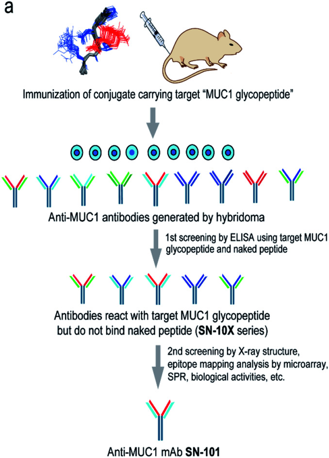
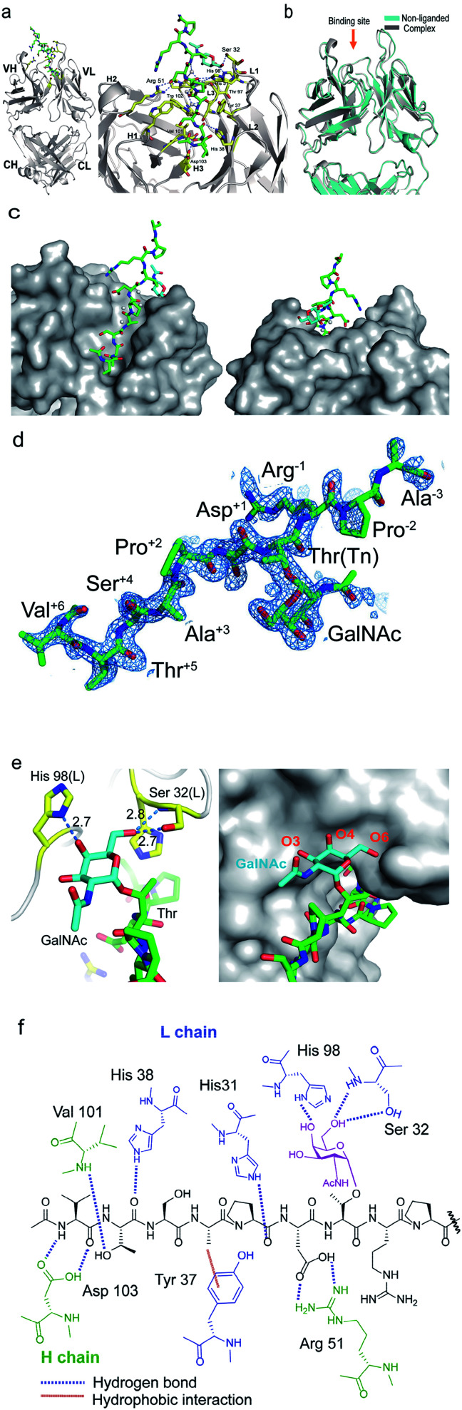
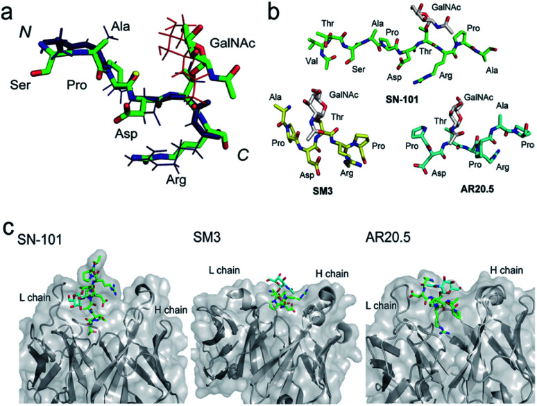
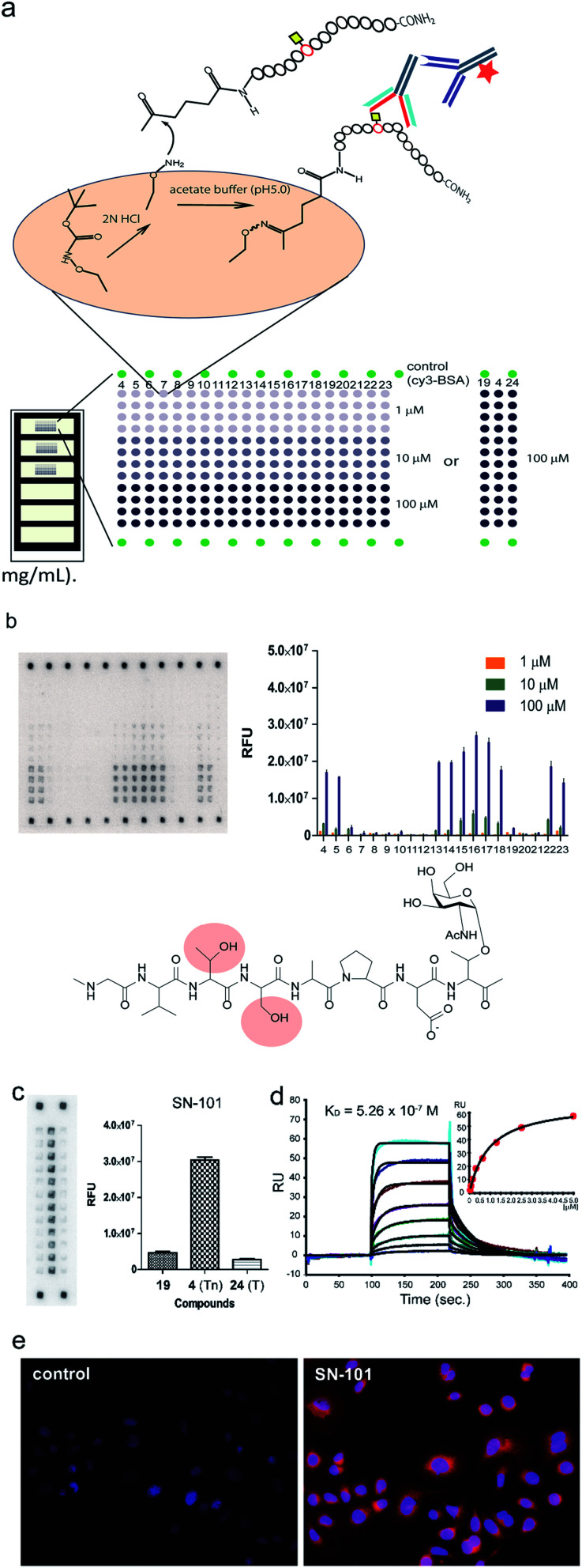
Aberrantly truncated immature O-glycosylation in proteins occurs in essentially all types of epithelial cancer cells, which was demonstrated to be a common feature of most adenocarcinomas and strongly associated with cancer proliferation and metastasis. Although extensive efforts have been made toward the development of anticancer antibodies targeting MUC1, one of the most studied mucins having cancer-relevant immature O-glycans, no anti-MUC1 antibody recognises carbohydrates and the proximal MUC1 peptide region, concurrently. Here we present a general strategy that allows for the creation of antibodies interacting specifically with glycopeptidic neoepitopes by using homogeneous synthetic MUC1 glycopeptides designed for the streamlined process of immunization, antibody screening, three-dimensional structure analysis, epitope mapping and biochemical analysis. The X-ray crystal structure of the anti-MUC1 monoclonal antibody SN-101 complexed with the antigenic glycopeptide provides for the first time evidence that SN-101 recognises specifically the essential epitope by forming multiple hydrogen bonds both with the proximal peptide and GalNAc linked to the threonine residue, concurrently. Remarkably, the structure of the MUC1 glycopeptide in complex with SN-101 is identical to its solution NMR structure, an extended conformation induced by site-specific glycosylation. We demonstrate that this method accelerates dramatically the development of a new class of designated antibodies targeting a variety of "dynamic neoepitopes" elaborated by disease-specific O-glycosylation in the immunodominant mucin domains and mucin-like sequences found in intrinsically disordered regions of many proteins.


Conflict of interest statement

There are no conflicts to declare.

Figures

Fig. 1
Fig. 1. Generation of epitope-defined anti-MUC1 antibodies. (a) A strategy for the generation of antibodies targeting glycopeptidic epitopes by using synthetic glycopeptides designed for the streamlined process from the immunization of “conformational glycopeptidic neoepitopes”, antibody selection, and characterization. (b) A list of compounds used in this study. Compound 1 was conjugated with KLH by using the Cys residue (red) or aminooxy-functionalized nanoparticles by using the ketone linker (blue) and used for the immunization. The first screening was performed by ELISA immobilizing compounds 1 and 2 using Cys residue (red) to collect antibodies binding selectively with glycopeptide 1. Compound 3 was used for the co-crystallization with SN-101. Compounds 4–24 were displayed on the microarray by means of the ketone linker (blue) and employed for epitope mapping analysis. Compound 25 was used for the SPR analysis by immobilizing with Cys residue (red).
Fig. 2
Fig. 2. Crystal structures of SN-101 Fab and its complex with the MUC1 glycopeptide 3 (PDB: 6KX0 and 6KX1). (a) Overall structure (left) and an enlarged picture focusing on the binding site (right) of SN-101 Fab complexed with 3. (b) Superposition of two SN-101 Fab structures of the complex (blue) and non-liganded (gray) forms. (c) Surface representations of SN-101 in complex with 3 (gray), seen from the back and front. Glycopeptide 3 is shown as a stick model with carbon atoms in green. (d) 2FoFc electron density map represents 10 amino acid residues of 15 mer MUC1 glycopeptide 3 bound to SN-101. (e) 3D structures focusing on GalNAc recognition by SN-101. Binding interaction is mediated by hydrogen bonds between O-4 and O-6 of GalNAc (blue) with His98 and Ser32 of the light chain (yellow), respectively (left). SN-101 surface represents the topology of the GalNAc binding site (left). (f) Schematic image of the interactions between SN-101 Fab and MUC1 glycopeptide 3.
Fig. 3
Fig. 3. SN-101 recognises the specifically dynamic glycopeptidic neoepitope. (a) Superposition of the solution NMR structure of 23 mer MUC1 glycopeptide [Ac-Gly-Val-Thr-Ser-Ala-Pro-Asp-Thr(Tn)-Arg-Pro-Ala-Pro-Gly-Ser-Thr-Ala-Pro-Pro-His-Gly-Val-Thr-NH2] and the X-ray crystal structure of 15 mer MUC1 glycopeptide 3 complexed with SN-101. 30 lowest energy NMR structures in the Ser-Ala-Pro-Asp-Thr(Tn)-Arg region was overlaid. Blue stick represents the peptide backbone of the NMR structure. (b) Conformations of Tn-glycosylated MUC fragments bound to SN-101, SM3, and AR20.5. (c) Comparison of the X-ray crystal structures of SN-101 (this study, PDB: 6KX1), SM3 (PDB: 5A2K), and AR20.5 (PDB: 5T78) in complex with Tn-glycosylated MUC1 fragments.
Fig. 4
Fig. 4. Interaction of SN-101 with synthetic MUC1 glycopeptides and membrane bound MUC1 of human breast cancer cells. (a) Preparation and layout of the microarray displaying MUC1 peptide and glycopeptides. (b) Epitope mapping analysis of SN-101 using a microarray displaying compounds 4–24. Compounds 4–12 are glycopeptides whose N-terminal residues decrease one by one. Similarly, compounds 13–18 are the glycopeptides whose C-terminal residues increase one by one. A naked MUC1 peptide 19, MUC1 fragments having two Tn antigens 20–23, and compound 24 with the T antigen at immunodominant Pro-Asp-Thr-Arg are also used. The results represent the average fluorescence intensities of the spots (n = 4) for all compounds in each concentration (1, 10, and 100 μM). The chemical structure shows an essential epitope for SN-101 and glycosylation at the highlighted Thr or Ser residue abrogates the antibody recognition. (c) Glycan specific binding of SN-101 revealed by glycoform-focused microarray prepared from 100 μM MUC1 glycopeptides 4, 19, and 24. The results represent the average fluorescence intensities of spots (n = 12) for compounds 19 (naked), 4 (Tn antigen), and 24 (T antigen), respectively. (d) Binding affinity of SN-101 Fab to a MUC1 glycopeptide 25 determined from the SPR binding curve (KD = 5.26 × 10⁻⁷ M). (e) Interaction of SN-101 with cancer cell surface MUC1 illuminated by fluorescence-labelled anti-mouse IgG mAb.

Similar articles

Cited by

References

    1. Cheever M. A. et al. . Clin. Cancer Res. 2009;15:5323–5337. doi: 10.1158/1078-0432.CCR-09-0737. - DOI - PMC - PubMed
    1. Nath S. Mukherjee P. Trends Mol. Med. 2014;20:332–342. doi: 10.1016/j.molmed.2014.02.007. - DOI - PMC - PubMed
    1. Kudelka M. R. et al. . Adv. Cancer Res. 2015;126:53–135. doi: 10.1016/bs.acr.2014.11.002. - DOI - PMC - PubMed
    1. Hollingsworth M. A. Swanson B. J. Nat. Rev. Cancer. 2004;4:45–60. doi: 10.1038/nrc1251. - DOI - PubMed
    1. Hattrup C. L. Gendler S. J. Annu. Rev. Physiol. 2007;70:431–457. doi: 10.1146/annurev.physiol.70.113006.100659. - DOI - PubMed