. 2019 Nov 22;11(3):699-712.
doi: 10.1039/c9sc04831f.

A complete picture of protein unfolding and refolding in surfactants

Affiliations

A complete picture of protein unfolding and refolding in surfactants

Jannik Nedergaard Pedersen et al. Chem Sci. .

Abstract

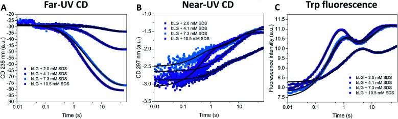
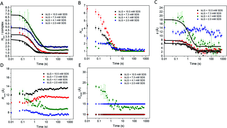
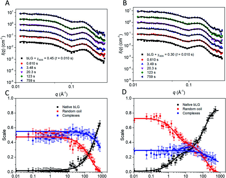
Interactions between proteins and surfactants are of relevance in many applications including food, washing powder formulations, and drug formulation. The anionic surfactant sodium dodecyl sulfate (SDS) is known to unfold globular proteins, while the non-ionic surfactant octaethyleneglycol monododecyl ether (C12E8) can be used to refold proteins from their SDS-denatured state. While unfolding have been studied in detail at the protein level, a complete picture of the interplay between protein and surfactant in these processes is lacking. This gap in our knowledge is addressed in the current work, using the β-sheet-rich globular protein β-lactoglobulin (bLG). We combined stopped-flow time-resolved SAXS, fluorescence, and circular dichroism, respectively, to provide an unprecedented in-depth picture of the different steps involved in both protein unfolding and refolding in the presence of SDS and C12E8. During unfolding, core-shell bLG-SDS complexes were formed within ∼10 ms. This involved an initial rapid process where protein and SDS formed aggregates, followed by two slower processes, where the complexes first disaggregated into single protein structures situated asymmetrically on the SDS micelles, followed by isotropic redistribution of the protein. Refolding kinetics (>100 s) were slower than unfolding (<30 s), and involved rearrangements within the mixing deadtime (∼5 ms) and transient accumulation of unfolded monomeric protein, differing in structure from the original bLG-SDS structure. Refolding of bLG involved two steps: extraction of most of the SDS from the complexes followed by protein refolding. These results reveal that surfactant-mediated unfolding and refolding of proteins are complex processes with rearrangements occurring on time scales from sub-milliseconds to minutes.


Conflict of interest statement

There are no conflicts to declare.

Figures

Fig. 1
Fig. 1. (A) Stick representation of octaethylene glycol monododecyl ether (C12E8) and sodium dodecyl sulfate (SDS). (B) Crystal structure of bLG (PDB entry 3NPO). (C) Small-angle X-ray scattering data of β-lactoglobulin (bLG), SDS, C12E8, and C12E8-SDS mixed micelles as well as model fits to bLG, SDS, and C12E8 data.
Fig. 2
Fig. 2. Unfolding kinetics of bLG followed by mixing bLG with different concentrations of SDS and tracked with (A) far-UV CD at 235 nm, (B) near-UV CD at 297 nm, and (C) fluorescence at 355 nm. Fits with exponential decay functions are plotted and the corresponding half times (t1/2) from fitting are shown in Table 1.
Fig. 3
Fig. 3. (A) Stopped-flow SAXS data of bLG mixed with 10.5 mM SDS at a time resolution of ∼4 ms and measured for 12.5 min. The arrows show the progression over time. (B) The model used for analysis of the SAXS data for unfolding of bLG mixed with SDS with Rcore representing the core radius, ε representing the axis ratio, Dhead the head group thickness, and an adjustable core offset represented by s. The model is based on Pilz et al.
Fig. 4
Fig. 4. Stopped-flow SAXS data of bLG mixed with (A) 10.5 mM SDS, (B) 7.3 mM SDS, (C) 4.1 mM SDS, and (D) 2.0 mM SDS at selected times through the series. Lines represent fits to the core–shell model with an adjustable core offset. For clarity, the data has been scaled with a factor of 3 for every displayed time step.
Fig. 5
Fig. 5. Structural model parameters obtained from fitting the core–shell model with an adjustable core offset to the stopped-flow unfolding SAXS data. The obtained parameters are: (A) number of proteins per complex, (B) number of micelles per complex, (C) core displacement, (D) radius of the core, and (E) size of the head group. For (A and C), single exponential decay functions were fitted to the data and the half times (t1/2) are displayed in Table 1. The core displacement at 2.0 mM SDS could not be fitted with an exponential function.
Fig. 6
Fig. 6. Refolding kinetics followed by mixing bLG-SDS complexes with two different mol ratios of C12E8 tracked with: (A) far-UV CD at 235 nm, (B) near-UV CD at 297 nm, and (C) fluorescence at 355 nm. Fits with exponential decay functions are plotted and the corresponding half times (t1/2) from fitting are shown in Table 1.
Fig. 7
Fig. 7. Stopped-flow SAXS data of bLG-SDS complexes mixed with C12E8 to (A) χSDS = 0.45 and (B) χSDS = 0.30. For clarity, the data has been scaled with a factor of 3 for every displayed time step. Contributions from the scales of the various protein species for data with (C) χSDS = 0.45 and (D) χSDS = 0.30. In (C and D) exponential fits are plotted and the corresponding half times (t1/2) from fitting are shown in Table 1.
Fig. 8
Fig. 8. Sketch showing the suggested unfolding and refolding scheme when bLG is unfolded with SDS and subsequently refolded with C12E8. Unfolding occurs in three steps with an initial clustering of SDS and protein within the deadtime of the experiment, followed by disaggregation of the complexes into individual SDS micelles with asymmetrically distributed protein, as seen by changes in Trp fluorescence, near-UV CD, and SAXS. In the last step, the protein is redistributed more symmetrically around the SDS micelle, as seen with changes in SAXS, near/far-UV CD, and Trp fluorescence. Refolding is somewhat more complex and slow and also involves three steps. Within the deadtime, a fraction of the protein dissociates from the surfactant micelles while the rest remains in complex, as observed by SAXS. Next, the unfolded protein is folded to a native state and finally the protein still in complex dissociates and refolds. α-Helices are shown in red and two α-helices represent a structure with native-like secondary structure, while three helices represent structures with increased α-helical content. Note that for the refolding process there are additional changes in aggregation state of some of the species, which for simplicity are not indicated in the figure.

References

    1. Kirk O. Borchert T. V. Fuglsang C. C. Industrial enzyme applications. Curr. Opin. Biotechnol. 2002;13:345–351. doi: 10.1016/S0958-1669(02)00328-2. - DOI - PubMed
    1. Otzen D. E. Protein-surfactant interactions: a tale of many states. Biochim. Biophys. Acta. 2011;1814:562–591. doi: 10.1016/j.bbapap.2011.03.003. - DOI - PubMed
    1. Otzen D. E. Proteins in a brave new surfactant world. Curr. Opin. Colloid Interface Sci. 2015;20:161–169. doi: 10.1016/j.cocis.2015.07.003. - DOI
    1. Nielsen M. M. Andersen K. K. Westh P. Otzen D. E. Unfolding of beta-sheet proteins in SDS. Biophys. J. 2007;92:3674–3685. doi: 10.1529/biophysj.106.101238. - DOI - PMC - PubMed
    1. Andersen K. K. Oliveira C. L. Larsen K. L. Poulsen F. M. Callisen T. H. Westh P. Pedersen J. S. Otzen D. The role of decorated SDS micelles in sub-CMC protein denaturation and association. J. Mol. Biol. 2009;391:207–226. doi: 10.1016/j.jmb.2009.06.019. - DOI - PubMed