. 2020 Jan 8;11(7):1878-1891.
doi: 10.1039/c9sc05094a.

Chemical tagging for sensitive determination of uridine modifications in RNA

Affiliations

Chemical tagging for sensitive determination of uridine modifications in RNA

Qing-Yun Cheng et al. Chem Sci. .

Abstract

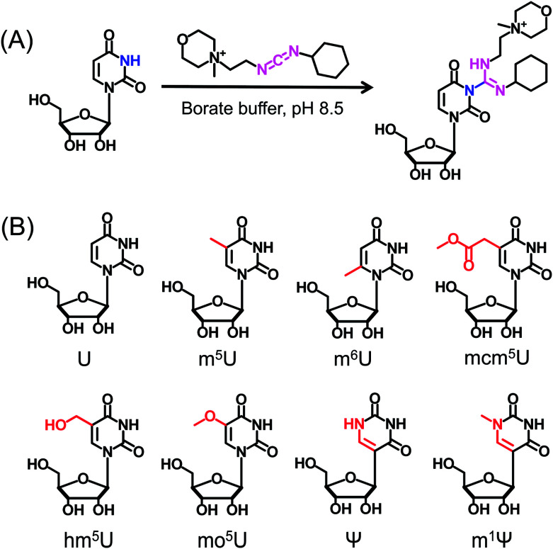
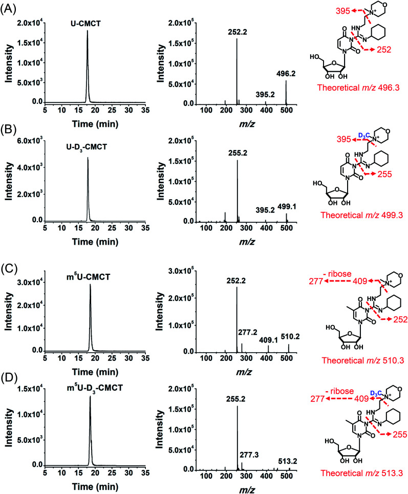
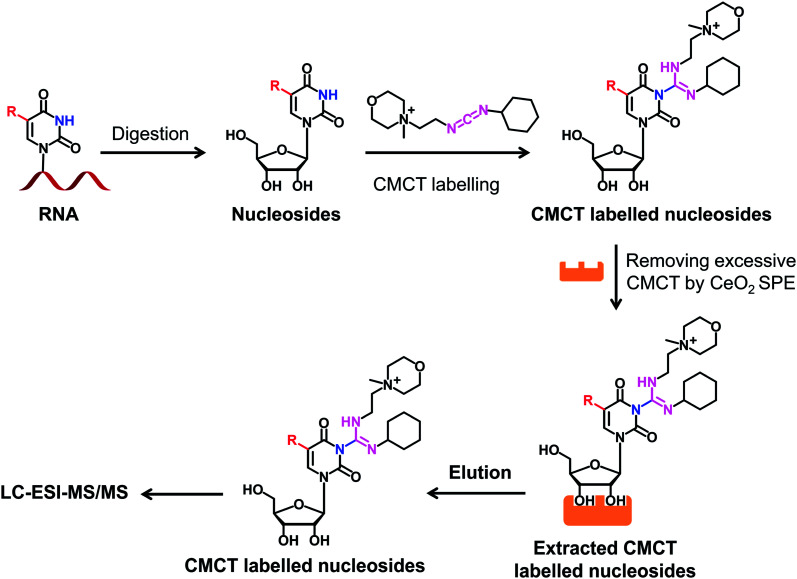
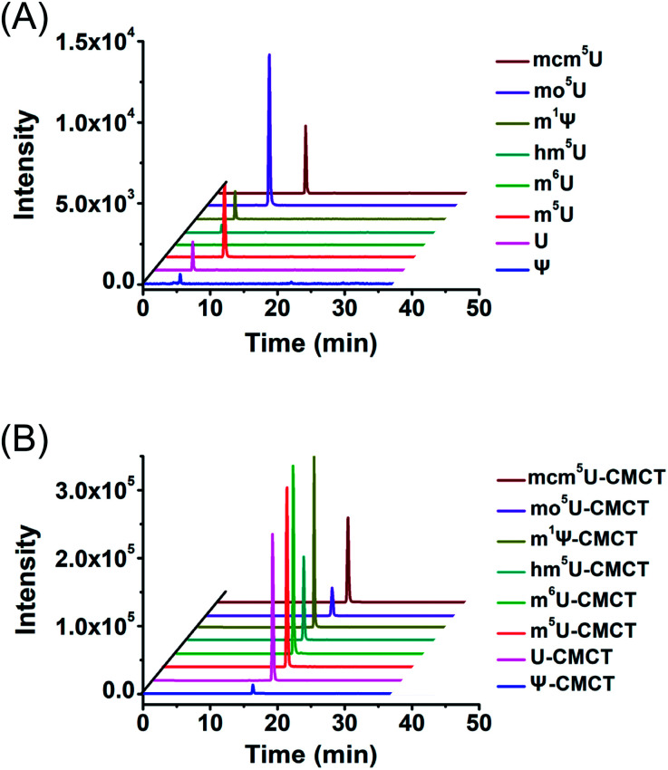
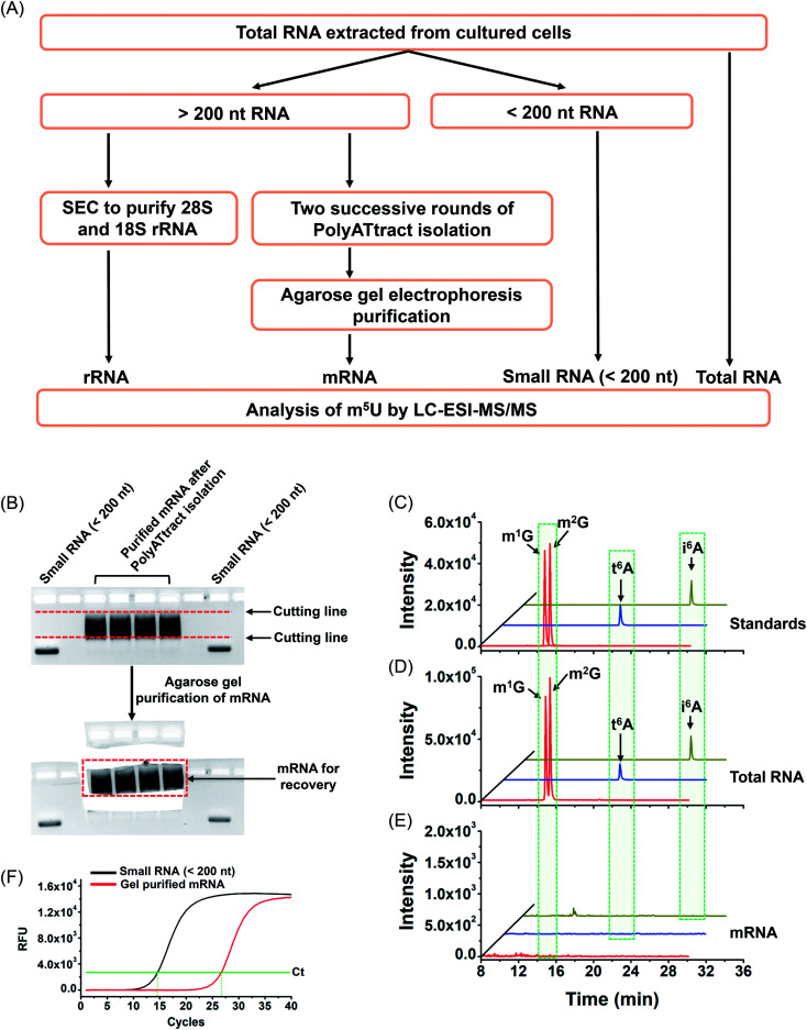
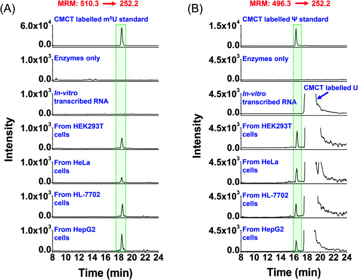
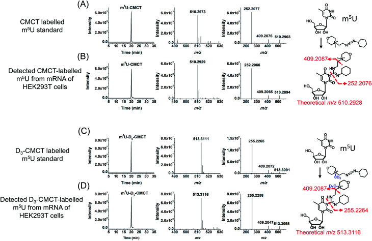
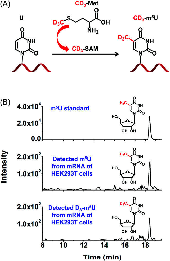
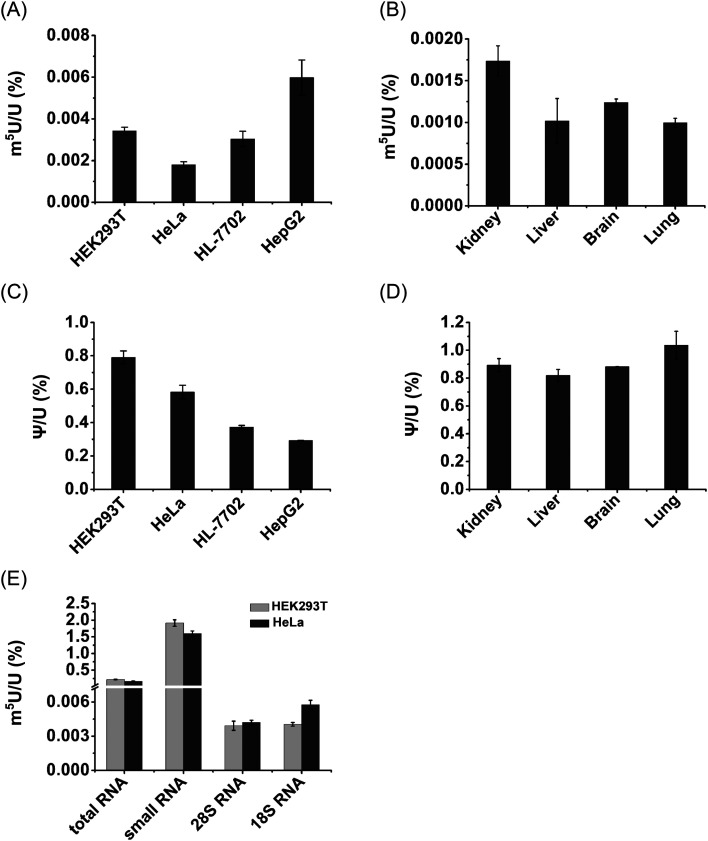
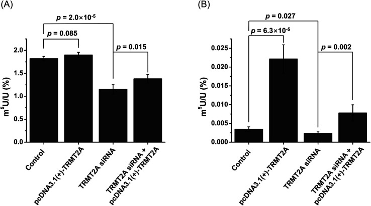
The discovery of dynamic and reversible modifications in messenger RNA (mRNA) is opening new directions in RNA modification-mediated regulation of biological processes. Methylation is the most prevalent modification occurring in mRNA and the methyl group is mainly decorated in the adenine, cytosine, and guanine base or in the 2'-hydroxyl group of ribose. However, methylation of the uracil base (5-methyluridine, m5U) has not been discovered in mRNA of eukaryotes. In the current study, we established a method of N-cyclohexyl-N'-β-(4-methylmorpholinium) ethylcarbodiimide p-toluenesulfonate (CMCT) labelling coupled with liquid chromatography-electrospray ionization-mass spectrometry (LC-ESI-MS/MS) analysis for the sensitive determination of uridine modifications in RNA. Our results demonstrated that the detection sensitivities of uridine modifications in RNA increased up to 1408 fold upon CMCT labelling. Using the developed method, we identified the distinct existence of m5U in mRNA of various mammalian cells and tissues. In addition, the stable isotope tracing monitored by mass spectrometry revealed that the methyl group of m5U originated from S-adenosyl-l-methionine (SAM). Our study expanded the list of modifications occurring in mRNA of mammals. Future work on transcriptome-wide mapping of m5U will further uncover the functional roles of m5U in mRNA of mammals.


Conflict of interest statement

The authors declare no competing financial interest.

Figures

Fig. 1
Fig. 1. Chemical reaction for labelling of uridine with CMCT. (A) CMCT carries a carbodiimide group that can selectively react with the NH group at the N3 position of the uracil base. (B) The chemical structures of uridine and uridine modifications.
Fig. 2
Fig. 2. Examination of the CMCT/D3-CMCT labelling reaction. Extracted-ion chromatogram and fragment ions of (A) CMCT labelled U, (B) D3-CMCT labelled U, (C) CMCT labelled m5U, and (D) D3-CMCT labelled m5U. The mass transitions 496.3 → 252.2 for CMCT labelled U, 499.3 → 255.2 for D3-CMCT labelled U, 510.3 → 252.2 for CMCT labelled m5U, and 513.3 → 255.2 for D3-CMCT labelled m5U were used for the detection in MRM detection mode.
Fig. 3
Fig. 3. Schematic illustration of the procedure for determination of uridine modifications by CMCT labelling coupled with LC-ESI-MS/MS analysis. Isolated RNA was enzymatically digested followed by CMCT labelling. Then the CMCT labelled uridine modifications were extracted and enriched by CeO2 solid phase extraction (SPE). The resulting CMCT labelled uridine modifications were subjected to LC-ESI-MS/MS analysis.
Fig. 4
Fig. 4. Extracted-ion chromatograms of Ψ, U, m5U, m6U, mcm5U, hm5U, m1Ψ and mo5U without (A) and with (B) CMCT labelling. The optimized MS conditions are listed in Table S1 in the ESI.
Fig. 5
Fig. 5. Schematic illustration of the extraction of different species of RNA and evaluation of the purity of the isolated mRNA. (A) Schematic illustration of the extraction of different species of RNA. (B) Agarose gel electrophoresis-based purification. (C) Extracted-ion chromatograms of the standards of 4 modifications (m1G, m2G, t6A, and i6A). (D) Extracted-ion chromatograms of the 4 modifications detected in total RNA of HEK293T cells. (E) Extracted-ion chromatograms of the 4 modifications detected in mRNA of HEK293T cells. (F) Evaluation of the purity of the isolated mRNA by real-time quantitative PCR.
Fig. 6
Fig. 6. Extracted-ion chromatograms of the detected m5U and Ψ in mRNA of various human cells. (A) Extracted-ion chromatograms of m5U standards and the m5U detected in mRNA from human HEK293T, HeLa, HL-7702, and HepG2 cells. (B) Extracted-ion chromatograms of Ψ standards and the Ψ detected in mRNA from human HEK293T, HeLa, HL-7702, and HepG2 cells. “Enzyme only” represents the sample only containing the enzymes used for digestion (S1 nuclease, venom phosphodiesterase I, and calf intestinal alkaline phosphatase) and omitting the mRNA. The sequence of the in vitro transcribed RNA can be found in Table S7 in the ESI. The mass transitions 510.3 → 252.2 for CMCT labelled m5U and 496.3 → 252.2 for CMCT labelled Ψ were used for the detection in MRM detection mode.
Fig. 7
Fig. 7. Identification of m5U in mRNA of HEK293T cells by high-resolution mass spectrometry analysis. (A) The extracted-ion chromatogram (left panel), MS spectrum (middle panel) and MS/MS spectrum (right panel) of the CMCT labelled m5U standard. (B) The extracted-ion chromatogram (left panel), MS spectrum (middle panel) and MS/MS spectrum (right panel) of CMCT labelled m5U from mRNA of HEK293T cells. (C) The extracted-ion chromatogram (left panel), MS spectrum (middle panel) and MS/MS spectrum (right panel) of the D3-CMCT labelled m5U standard. (D) The extracted-ion chromatogram (left panel), MS spectrum (middle panel) and MS/MS spectrum (right panel) of D3-CMCT labelled m5U from mRNA of HEK293T cells.
Fig. 8
Fig. 8. Identification of m5U in mRNA of HEK293T cells using stable isotope tracing monitored by mass spectrometry. (A) DMEM medium supplied with D3-Met was used for the cell culturing. D3-Met could be converted to D3-SAM that is a methylating reagent for methylation of nucleic acids. (B) The extracted-ion chromatograms of the CMCT labelled m5U standard (upper panel), detected CMCT labelled m5U from mRNA of HEK293T cells (middle panel), and detected CMCT labelled D3-m5U from mRNA of HEK293T cells (bottom panel).
Fig. 9
Fig. 9. Measured contents of m5U in different cells, tissues and RNA species. (A) Measured contents of m5U in mRNA from various human cell lines. (B) Measured contents of m5U in mRNA from various mouse tissues. (C) Measured contents of Ψ in mRNA from various human cell lines. (D) Measured contents of Ψ in mRNA from various mouse tissues. (E) Measured contents of m5U in different RNA species from HEK293T and HeLa cells. These data represent the mean and standard deviation of results acquired from three independent experiments.
Fig. 10
Fig. 10. Effect of overexpression and siRNA knockdown of TRMT2A on the contents of m5U in small RNA and mRNA. (A) Contents of m5U in small RNA upon overexpression or siRNA knockdown of TRMT2A. (B) Contents of m5U in mRNA upon overexpression or siRNA knockdown of TRMT2A. Two-side unpaired t-test was performed. These data represent the mean and standard deviation of results acquired from three independent experiments.

References

    1. Chen K. Zhao B. S. He C. Cell Chem. Biol. 2016;23:74–85. doi: 10.1016/j.chembiol.2015.11.007. - DOI - PMC - PubMed
    1. Luo C. Hajkova P. Ecker J. R. Science. 2018;361:1336–1340. doi: 10.1126/science.aat6806. - DOI - PMC - PubMed
    1. Frye M. Harada B. T. Behm M. He C. Science. 2018;361:1346–1349. doi: 10.1126/science.aau1646. - DOI - PMC - PubMed
    1. Roundtree I. A. Evans M. E. Pan T. He C. Cell. 2017;169:1187–1200. doi: 10.1016/j.cell.2017.05.045. - DOI - PMC - PubMed
    1. Boccaletto P. Machnicka M. A. Purta E. Piatkowski P. Baginski B. Wirecki T. K. de Crecy-Lagard V. Ross R. Limbach P. A. Kotter A. Helm M. Bujnicki J. M. Nucleic Acids Res. 2018;46:D303–D307. doi: 10.1093/nar/gkx1030. - DOI - PMC - PubMed