

Cardiac hydatid cyst in the right ventricle - A rare case report of echinococcosis presentation
- PMID: 34123377
- PMCID: PMC8175272
- DOI: 10.1016/j.amsu.2021.102427
Cardiac hydatid cyst in the right ventricle - A rare case report of echinococcosis presentation
Abstract
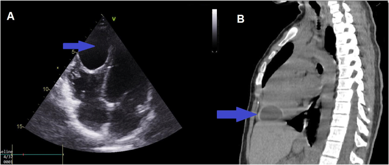
Introduction: The hydatid cyst (HC) of the right ventricle (RV) is an extremely uncommon and a serious location that can cause sudden death following pulmonary embolism, obstruction of the valvular orifice or anaphylactic shock.
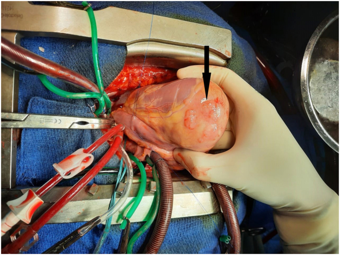
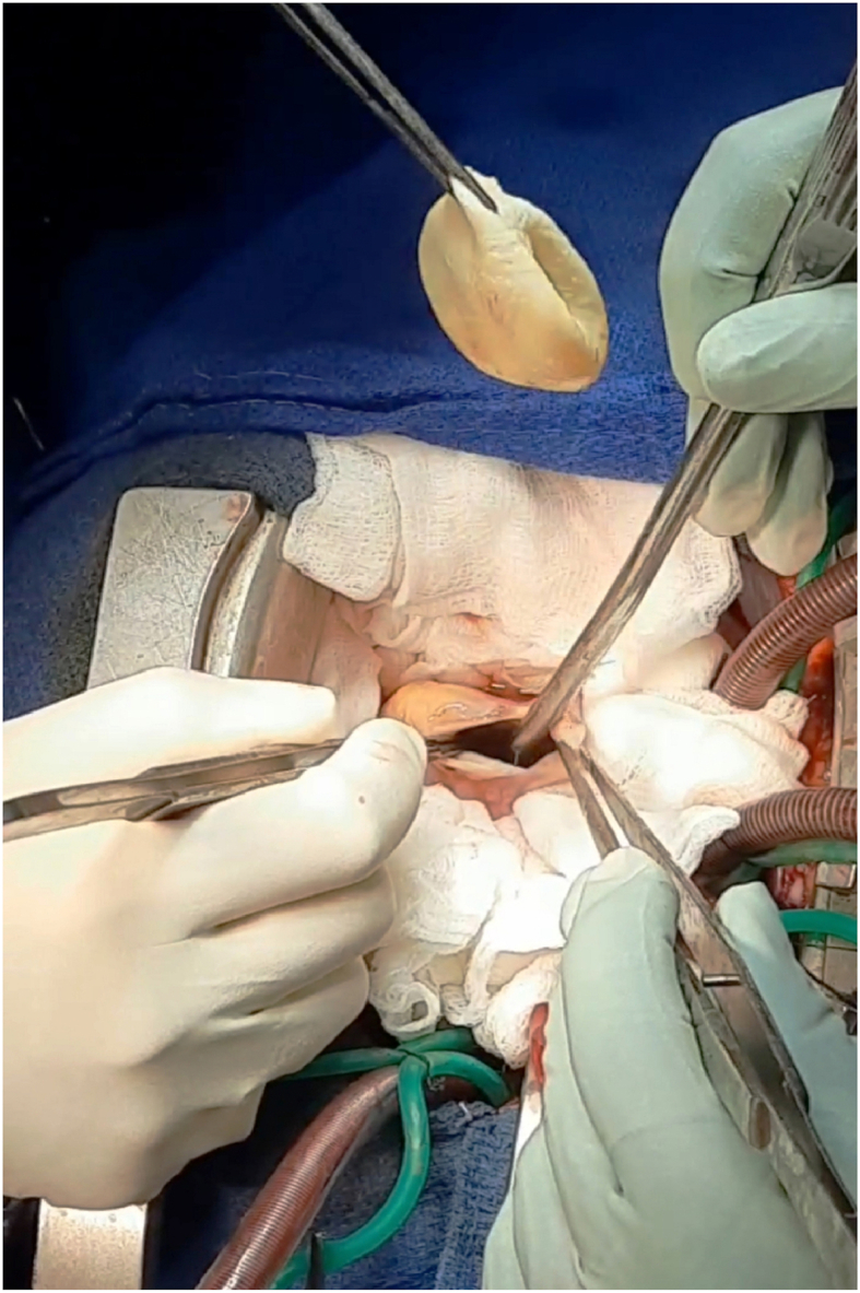
Case presentation: We report a case of a 14 years-old girl with a HC of the RV. Surgical excision of the HC under Cardiopulmonary bypass (CPB) was successful in managing this rare case.
Clinical discussion: Cardiac HC is extremely rare. It represents only 0.5-2% of all hydatid cases. However, RV location is very severe. It has a tendency to rupture intracavitarily and causes sudden death in 30% of cases. Its diagnosis is based on echocardiography, computed tomography scan and magnetic resonance imaging. The surgical treatment under CPB with anthelmintic therapy seems to improve the prognostic outcomes.
Conclusion: Cardiac HC must be always suspected in endemic countries, especially in patients with a family history of HC.
Keywords: Case report; Heart; Hydatid cyst; Right ventricle; SCARE guidelines.
© 2021 The Authors.
Conflict of interest statement
The authors declare no conflicts of interest.
Figures
References
-
- Blanton R. Echinococcosis. In: Behrman R.E., KliegmanRM, Jenson H.B., editors. Nelson Textbook of Pediatrics. seventeenth ed. WB Saunders Company; Philadelphia: 2004. pp. 1173–1174.
Publication types
LinkOut - more resources
Full Text Sources
