

Reflectance Confocal Microscopy of Aging Skin and Skin Cancer
- PMID: 34123564
- PMCID: PMC8172052
- DOI: 10.5826/dpc.1103a68
Reflectance Confocal Microscopy of Aging Skin and Skin Cancer
Abstract
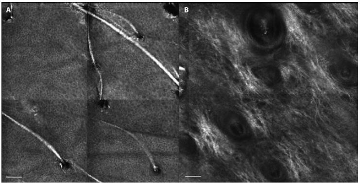
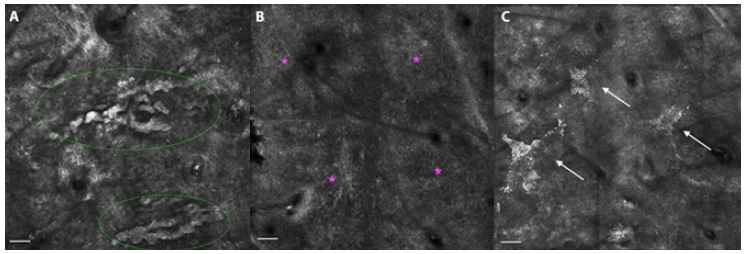
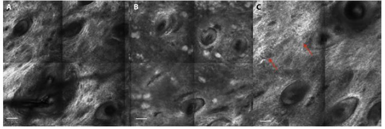
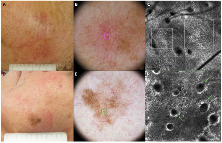
Skin aging is a complex process that causes morphologic variations. Some of these variations have been hypothesized to be involved in skin cancer development. This paper reviews current knowledge of the features of aged skin as seen with reflectance confocal microscopy (RCM). Basic principles of the technique are described, and the RCM features of healthy skin and skin cancer are briefly discussed. Moreover, the RCM features at different layers of young and elderly skin are described, as are the variations that occur with passing years and in relation to sun exposure that contribute to photoaging and the development of skin cancer. RCM enables the noninvasive evaluation, at quasi-histologic resolution, of aging-related skin changes, some of which are shared with skin cancer; this ability helps avoid skin biopsy. Further research is needed to understand the relation between skin aging and skin cancer development.
Keywords: Skin aging; blood vessels; collagen; reflectance confocal microscopy; skin cancer.
©2021 Guida et al.
Conflict of interest statement
Competing interests: The authors have no conflicts of interest to disclose.
Figures
Similar articles
-
Role of In Vivo Reflectance Confocal Microscopy in the Analysis of Melanocytic Lesions.Acta Dermatovenerol Croat. 2018 Apr;26(1):64-67. Acta Dermatovenerol Croat. 2018. PMID: 29782304 Review.
-
Introduction to reflectance confocal microscopy and its use in clinical practice.JAAD Case Rep. 2018 Nov 10;4(10):1014-1023. doi: 10.1016/j.jdcr.2018.09.019. eCollection 2018 Nov. JAAD Case Rep. 2018. PMID: 30456275 Free PMC article.
-
Using Reflectance Confocal Microscopy in Skin Cancer Diagnosis.Dermatol Clin. 2017 Oct;35(4):457-464. doi: 10.1016/j.det.2017.06.007. Epub 2017 Aug 7. Dermatol Clin. 2017. PMID: 28886802 Review.
-
Assessment of skin photoaging with reflectance confocal microscopy.Skin Res Technol. 2014 Aug;20(3):363-72. doi: 10.1111/srt.12127. Epub 2014 Feb 10. Skin Res Technol. 2014. PMID: 24506234 Clinical Trial.
-
In vivo assessment of chronological ageing and photoageing in forearm skin using reflectance confocal microscopy.Br J Dermatol. 2012 Aug;167(2):270-9. doi: 10.1111/j.1365-2133.2012.10943.x. Epub 2012 Jun 20. Br J Dermatol. 2012. PMID: 22428802
Cited by
-
Coffea arabica: An Emerging Active Ingredient in Dermato-Cosmetic Applications.Pharmaceuticals (Basel). 2025 Jan 27;18(2):171. doi: 10.3390/ph18020171. Pharmaceuticals (Basel). 2025. PMID: 40005985 Free PMC article. Review.
-
Non-invasive 3D imaging of human melanocytic lesions by combined ultrasound and photoacoustic tomography: a pilot study.Sci Rep. 2024 Feb 2;14(1):2768. doi: 10.1038/s41598-024-53220-y. Sci Rep. 2024. PMID: 38307985 Free PMC article.
-
Laser Treatment Monitoring with Reflectance Confocal Microscopy.Medicina (Kaunas). 2023 May 28;59(6):1039. doi: 10.3390/medicina59061039. Medicina (Kaunas). 2023. PMID: 37374244 Free PMC article.
-
Comparison of facial skin ageing in healthy Asian and Caucasian females quantified by in vivo line-field confocal optical coherence tomography 3D imaging.Skin Res Technol. 2024 Sep;30(9):e13643. doi: 10.1111/srt.13643. Skin Res Technol. 2024. PMID: 39221700 Free PMC article.
-
The effect of COVID-19 on surgical management of skin cancers of the head, face and neck in elderly patients.Skin Health Dis. 2023 Mar 28;3(2):e175. doi: 10.1002/ski2.175. eCollection 2023 Apr. Skin Health Dis. 2023. PMID: 37025367 Free PMC article.
References
Publication types
LinkOut - more resources
Full Text Sources