

Can Dermoscopy Be Used to Predict if a Melanoma Is In Situ or Invasive?
- PMID: 34123569
- PMCID: PMC8172039
- DOI: 10.5826/dpc.1103a79
Can Dermoscopy Be Used to Predict if a Melanoma Is In Situ or Invasive?
Abstract
Background: The preoperative prediction of whether melanomas are invasive or in situ can influence initial management.
Objectives: This study evaluated the accuracy rate, interobserver concordance, sensitivity and specificity in determining if a melanoma is invasive or in situ, as well as the ability to predict invasive melanoma thickness based on clinical and dermoscopic images.
Methods: In this retrospective, single-center investigation, 7 dermatologists independently reviewed clinical and dermoscopic images of melanomas to predict if they were invasive or in situ and, if invasive, their Breslow thickness. Fleiss' and Cohen's kappa (κ) were used for interobserver concordance and agreement with histopathological diagnosis.
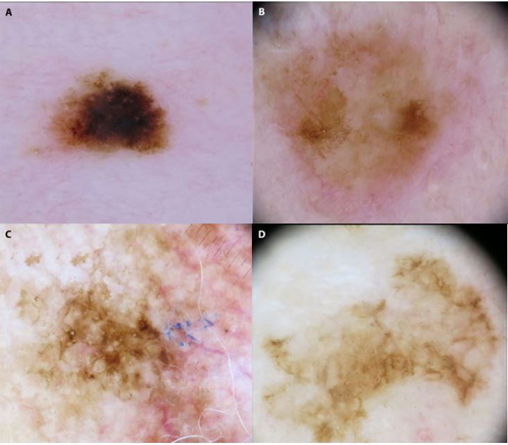
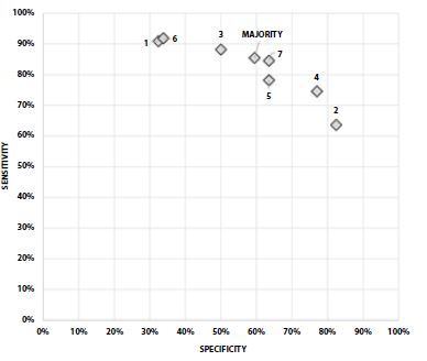
Results: We included 184 melanomas (110 invasive and 74 in situ). Diagnostic accuracy ranged from 67.4% to 76.1%. Accuracy rates for in situ and invasive melanomas were 57.5% (95% confidence interval [CI], 53.1%-61.8%) and 81.7% (95% CI, 78.8%-84.4%), respectively. Interobserver concordance was moderate (κ = 0.47; 95% CI, 0.44-0.51). Sensitivity for predicting invasiveness ranged from 63.6% to 91.8% for 7 observers, while specificity was 32.4%-82.4%. For all correctly predicted invasive melanomas, agreement between predictions and correct thickness over or under 1.0 mm was moderate (κ = 0.52; 95% CI, 0.45-0.58). All invasive melanomas incorrectly predicted by any observer as in situ had a thickness <1.0 mm. All 32 melanomas >1.0 mm were correctly predicted to be invasive by all observers.
Conclusions: Accuracy rates for predicting thick melanomas were excellent, melanomas inaccurately predicted as in situ were all thin, and interobserver concordance for predicting in situ or invasive melanomas was moderate. Preoperative dermoscopy of suspected melanomas is recommended for choosing appropriate surgical margins.
Keywords: Breslow thickness; Dermoscopy; Inter Observer Variability; Melanoma; Projections and Predictions.
©2021 Polesie et al.
Conflict of interest statement
Competing interests: The authors have no conflicts of interest to disclose.
Figures
Similar articles
-
Multispectral Imaging Algorithm Predicts Breslow Thickness of Melanoma.J Clin Med. 2021 Dec 30;11(1):189. doi: 10.3390/jcm11010189. J Clin Med. 2021. PMID: 35011930 Free PMC article.
-
Interobserver Agreement on Dermoscopic Features and their Associations with In Situ and Invasive Cutaneous Melanomas.Acta Derm Venereol. 2021 Oct 14;101(10):adv00570. doi: 10.2340/actadv.v101.281. Acta Derm Venereol. 2021. PMID: 34596231 Free PMC article.
-
Assessment of melanoma thickness based on dermoscopy images: an open, web-based, international, diagnostic study.J Eur Acad Dermatol Venereol. 2022 Nov;36(11):2002-2007. doi: 10.1111/jdv.18436. Epub 2022 Jul 26. J Eur Acad Dermatol Venereol. 2022. PMID: 35841304 Free PMC article.
-
Dermoscopic evaluation of superficial spreading melanoma.An Bras Dermatol. 2021 Mar-Apr;96(2):139-147. doi: 10.1016/j.abd.2020.06.012. Epub 2021 Feb 1. An Bras Dermatol. 2021. PMID: 33637398 Free PMC article.
-
Dermoscopic features of thin melanomas: a comparative study of melanoma in situ and invasive melanomas smaller than or equal to 1mm.An Bras Dermatol. 2013 Sep-Oct;88(5):712-7. doi: 10.1590/abd1806-4841.20132017. An Bras Dermatol. 2013. PMID: 24173175 Free PMC article.
Cited by
-
Multispectral Imaging Algorithm Predicts Breslow Thickness of Melanoma.J Clin Med. 2021 Dec 30;11(1):189. doi: 10.3390/jcm11010189. J Clin Med. 2021. PMID: 35011930 Free PMC article.
-
Skin Cancer Diagnosis by Lesion, Physician, and Examination Type: A Systematic Review and Meta-Analysis.JAMA Dermatol. 2025 Feb 1;161(2):135-146. doi: 10.1001/jamadermatol.2024.4382. JAMA Dermatol. 2025. PMID: 39535756
-
Improving Skin Cancer Diagnostics Through a Mobile App With a Large Interactive Image Repository: Randomized Controlled Trial.JMIR Dermatol. 2023 Aug 9;6:e48357. doi: 10.2196/48357. JMIR Dermatol. 2023. PMID: 37624707 Free PMC article.
-
Evaluation of Melanoma Thickness with Clinical Close-up and Dermoscopic Images Using a Convolutional Neural Network.Acta Derm Venereol. 2022 Oct 11;102:adv00790. doi: 10.2340/actadv.v102.2681. Acta Derm Venereol. 2022. PMID: 36172695 Free PMC article.
-
Performance of a Machine Learning Algorithm on Lesions with a High Preoperative Suspicion of Invasive Melanoma.Acta Derm Venereol. 2024 Jul 18;104:adv40023. doi: 10.2340/actadv.v104.40023. Acta Derm Venereol. 2024. PMID: 39023145 Free PMC article. No abstract available.
References
-
- Pizzichetta MA, Argenziano G, Talamini R, et al. Dermoscopic criteria for melanoma in situ are similar to those for early invasive melanoma. Cancer. 2001;91(5):992–997. - PubMed
-
- Swedish guidelines for malignant melanoma [Article in Swedish] [Accessed: November 17, 2020]. Last updated April 29, 2019. https://kunskapsbanken.cancercentrum.se/globalassets/cancerdiagnoser/hud... .
-
- Garbe C, Amaral T, Peris K, et al. European Dermatology Form (EDF); European Association of Dermato-Oncology (EADO); European Organization for Research and Treatment of Cancer (EORTC) European consensus-based interdisciplinary guideline for melanoma. Part 2: Treatment - Update 2019. Eur J Cancer. 2020;126:159–177. doi: 10.1016/j.ejca.2019.11.015. - DOI - PubMed
Associated data
LinkOut - more resources
Full Text Sources