Biological assessment of a new ready-to-use hydraulic sealer
- PMID: 34123757
- PMCID: PMC8170373
- DOI: 10.5395/rde.2021.46.e21
Biological assessment of a new ready-to-use hydraulic sealer
Abstract
Objectives: This study compared the cytotoxicity, biocompatibility, and tenascin immunolabeling of a new ready-to-use hydraulic sealer (Bio-C Sealer) with MTA-Fillapex and white MTA-Angelus.
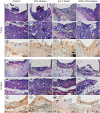
Materials and methods: L929 fibroblasts were cultivated and exposed to undiluted and diluted material extracts. Polyethylene tubes with or without (the control) the materials were implanted into the dorsa of rats. At 7 days and 30 days, the rats were euthanized, and the specimens were prepared for analysis; inflammation and immunolabeling were measured, and statistical analysis was performed (p < 0.05).
Results: MTA-Fillapex exhibited greater cytotoxicity than the other materials at all time points (p < 0.05). The undiluted Bio-C Sealer exhibited greater cytocompatibility at 6 and 48 hours than white MTA-Angelus, with higher cell viability than in the control (p < 0.05). White MTA-Angelus displayed higher cell viability than the control at 24 hours, and the one-half dilution displayed similar results at both 6 and 48 hours (p < 0.05). At 7 days and 30 days, the groups exhibited moderate inflammation with thick fibrous capsules and mild inflammation with thin fibrous capsules, respectively (p > 0.05). At 7 days, moderate to strong immunolabeling was observed (p > 0.05). After 30 days, the control and MTA-Fillapex groups exhibited strong immunolabeling, the white MTA-Angelus group exhibited moderate immunolabeling (p > 0.05), and the Bio-C Sealer group exhibited low-to-moderate immunolabeling, differing significantly from the control (p < 0.05).
Conclusions: Bio-C Sealer and white MTA-Angelus exhibited greater cytocompatibility than MTA-Fillapex; all materials displayed adequate biocompatibility and induced tenascin immunolabeling.
Keywords: Biocompatibility; Cytotoxicity; Hydraulic sealer; Mineral trioxide aggregate; Tenascin.
Copyright © 2021. The Korean Academy of Conservative Dentistry.
Conflict of interest statement
Conflict of Interest: No potential conflict of interest relevant to this article was reported.
Figures


References
-
- Vouzara T, Dimosiari G, Koulaouzidou EA, Economides N. Cytotoxicity of a new calcium silicate endodontic sealer. J Endod. 2018;44:849–852. - PubMed
-
- Shie MY, Chang HC, Ding SJ. Effects of altering the Si/Ca molar ratio of a calcium silicate cement on in vitro cell attachment. Int Endod J. 2012;45:337–345. - PubMed
-
- Han L, Okiji T. Uptake of calcium and silicon released from calcium silicate-based endodontic materials into root canal dentine. Int Endod J. 2011;44:1081–1087. - PubMed
-
- Reyes-Carmona JF, Felippe MS, Felippe WT. The biomineralization ability of mineral trioxide aggregate and Portland cement on dentin enhances the push-out strength. J Endod. 2010;36:286–291. - PubMed
LinkOut - more resources
Full Text Sources

