. 2021 May 27;6(22):14164-14173.
doi: 10.1021/acsomega.1c00741. eCollection 2021 Jun 8.

FRET-Based Genetically Encoded Sensor to Monitor Silver Ions

Affiliations

FRET-Based Genetically Encoded Sensor to Monitor Silver Ions

Neha Agrawal et al. ACS Omega. .

Abstract

Silver is commonly used in wound dressing, photography, health care products, laboratories, pharmacy, biomedical devices, and several industrial purposes. Silver (Ag+) ions are more toxic pollutants widely scattered in the open environment by natural processes and dispersed in soil, air, and water bodies. Ag+ binds with metallothionein, macroglobulins, and albumins, which may lead to the alteration of various enzymatic metabolic pathways. To analyze the uptake and metabolism of silver ions in vitro as well as in cells, a range of high-affinity fluorescence-based nanosensors has been constructed using a periplasmic protein CusF, a part of the CusCFBA efflux complex, which is involved in providing resistance against copper and silver ions in Escherichia coli. This nanosensor was constructed by combining of two fluorescent proteins (donor and acceptor) at the N- and C-terminus of the silver-binding protein (CusF), respectively. SenSil (WT) with a binding constant (K d) of 5.171 μM was more efficient than its mutant variants (H36D and F71W). This nanosensor allows monitoring the level of silver ions in real time in prokaryotes and eukaryotes without any disruption of cells or tissues.


Conflict of interest statement

The authors declare no competing financial interest.

Figures

Figure 1
Figure 1
(A) Construction of the FRET-based genetically encoded sensor for silver ions. (B) Crystal structure of CusF. (C) Energy transfer illustration in the absence (left) and presence (right) of the Ag+ ligand.
Figure 2
Figure 2
Spectral analysis of the FRET-based nanosensor. The emission spectrum was recorded after excitation of the sensor protein at 420 nm and recording the emission in the range of 450 to 600 nm both in the absence and presence of silver ions.
Figure 3
Figure 3
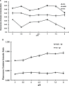
Buffer stability analysis of SenSil. (A) Emission intensity ratio was recorded in MOPS, phosphate-buffered saline (PBS), and Tris–Cl buffer with various ranges of pH values. The sensor was found to be stable in 20 mM MOPS buffer as least change in the FRET ratio was observed with this buffer. (B) Stability of the nanosensor was analyzed in MOPS buffer in a physiological pH range (5.0 to 8.0) in the absence and presence of 1 μM silver ions. Data are means of three independent replicates (n = 3). Vertical bars indicate the standard error.
Figure 4
Figure 4
In vitro specificity analysis of SenSil. (A) Specificity of the sensor protein was confirmed by measuring the FRET ratio in the presence of different metal ions (Ag+, Mn2+, Fe3+, Cu2+, and Ni2+). (B) Effect of essential metal ions such as NaCl, KCl, CaCl2, and MgCl2 on the efficiency of the nanosensor, which does not interfere with the specificity of SenSil. Data are means of three independent replicates (n = 3). Vertical bars indicate the standard error.
Figure 5
Figure 5
In vitro ligand-dependent FRET emission change of WT and mutant sensors in the presence of different silver ion concentrations. Affinity mutants H36D and F71W were created and compared with the WT sensor protein. Data are means of three independent replicates (n = 3). Vertical bars indicate the standard error.
Figure 6
Figure 6
Cell-based analysis of the nanosensor. FRET ratio changes in bacterial cells containing SenSil were recorded in response to 5 μM silver in a time-dependent manner. Data are means of three independent replicates (n = 3). Vertical bars indicate the standard error.
Figure 7
Figure 7
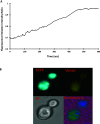
Real-time quantification of the dynamic flux rate change of Ag+ uptake in yeast cells. (A) The graph indicates change in the FRET ratio in the cytosol of yeast cells in the presence of 5 μM Ag+ along with the ratiometric changes in a single cell. Change in the FRET ratio indicates the import and binding of Ag+ ions with SenSil. (B) Confocal images showing cytosolic expression of SenSil in the presence of Ag+ ions in yeast (S. cerevisiae).
Figure 8
Figure 8
Real-time imaging of SenSil in the HEK-293T cell line. (A) HEK-293T cells were also transfected with SenSil and the emission intensity ratio changes were recorded for the defined period. The graph indicates the Venus/ECFP ratio change for 10 min. (B) Confocal images showing expression of SenSil in the presence of Ag+ in the HEK-293T cell line. (C) Cell viability studies of silver ions on HEK-293T. Viability of HEK-293T cells treated with increasing concentrations of Ag+ ions (0.01–20 μM) using the 3-[4,5-dimethylthiazol-2-yl]-2,5-diphenyltetrazolium bromide (MTT) assay. Percent cell viability was calculated with respect to the untreated control cells.

References

    1. Jaishankar M.; Tseten T.; Anbalagan N.; Mathew B. B.; Beeregowda K. N. Toxicity, mechanism and health effects of some heavy metals. Interdiscip. Toxicol. 2014, 7, 60–72. 10.2478/intox-2014-0009. - DOI - PMC - PubMed
    1. Kittleson J. T.; Loftin I. R.; Hausrath A. C.; Engelhardt K. P.; Rensing C.; McEvoy M. M. Periplasmic Metal-Resistance Protein CusF Exhibits High Affinity and Specificity for Both CuI and AgI. Biochemistry 2006, 45, 11096–11102. 10.1021/bi0612622. - DOI - PubMed
    1. Bury N. R.; McGeer J. C.; Wood C. M. Effects of altering freshwater chemistry on physiological responses of rainbow trout to silver exposure. Environ. Toxicol. Chem. 1999, 18, 49–55. 10.1002/etc.5620180107. - DOI
    1. Krizkova S.; Adam V.; Kizek R. Phytotoxicity of silver ions. Chem. Listy 2009, 103, 559–568.
    1. Fung M. C.; Bowen D. L. Silver products for medical indications: risk-benefit assessment. J. Toxicol., Clin. Toxicol. 1996, 34, 119–126. 10.3109/15563659609020246. - DOI - PubMed