

The expression and clinical prognostic value of protein phosphatase 1 catalytic subunit beta in pancreatic cancer
- PMID: 34125004
- PMCID: PMC8806868
- DOI: 10.1080/21655979.2021.1934243
The expression and clinical prognostic value of protein phosphatase 1 catalytic subunit beta in pancreatic cancer
Abstract
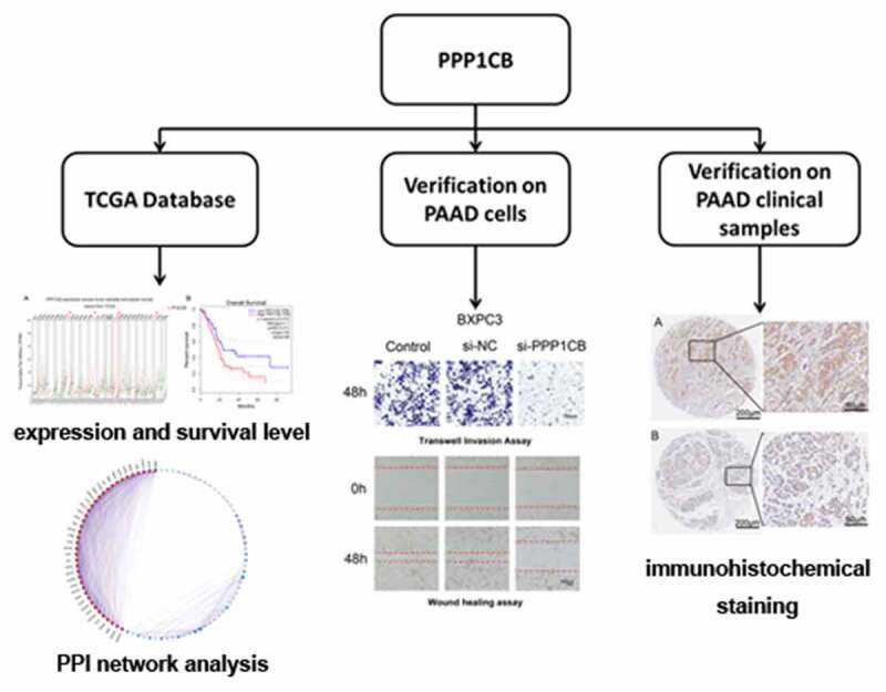
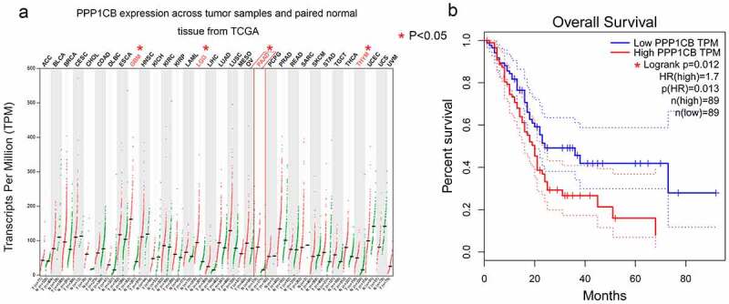
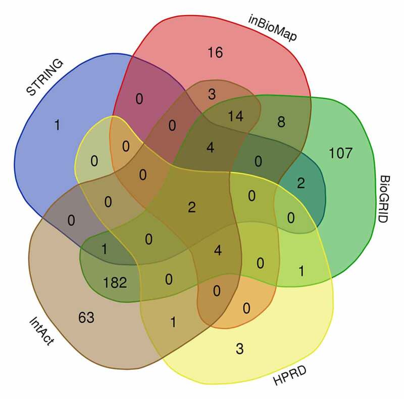
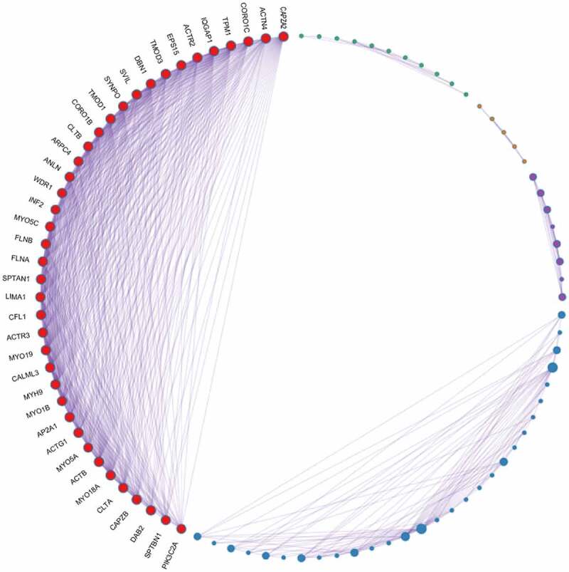
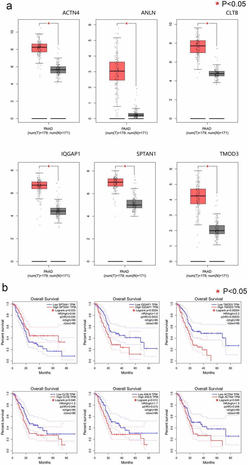
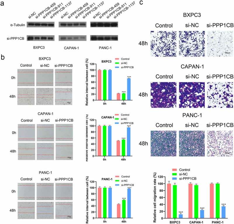
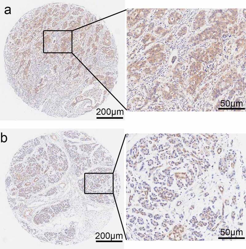
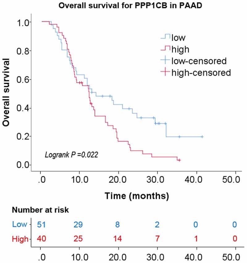
Pancreatic cancer (PAAD) is a common malignancy with a poor survival rate. The identification of novel biomarkers could improve clinical outcomes for patients with PAAD. Here we evaluated the expression and clinical significance of PPP1CB in PAAD. PPP1CB expression was higher in PAAD tissue than in matched paracancerous tissue (P < 0.05). We predicted a network of regulatory targets and protein interaction partners of PPP1CB, and identified a PPI network consisting of 39 node genes. The expression of 33 node genes was higher in PAAD tissue than in matching paracancerous tissue. High expression of the node genes ACTN4, ANLN, CLTB, IQGAP1, SPTAN1, and TMOD3 was associated with improved overall survival (P < 0.05). SiRNA knockdown of PPP1CB significantly reduced the migration and invasion of PAAD cells. A PPP1CB immunohistochemical staining was performed using a tissue microarray (TMA), consisting of tumor samples collected from 91 patients with PAAD (88 of which contained matched paracancerous tissues). The expression of PPP1CB in PAAD was significantly higher than in the matched paracancerous tissue, (P = 0.016). High PPP1CB expression was associated with patient sex (P = 0.048), alcohol use (P = 0.039), CEA (P= 0.038), N stage (P = 0.001), and invasion of nerve (P = 0.036). Furthermore, high PPP1CB expression was associated with significantly poorer overall survival (P = 0.022). Our data demonstrate that PPP1CB is associated with the migration and invasion of PAAD cells, and may be useful as an independent prognostic indicator for clinical outcome in patients with PAAD.
Keywords: Bioinformatics analysis; pancreatic adenocarcinoma; prognosis; protein phosphatase 1 catalytic subunit beta.
Figures
Similar articles
-
The up-regulation of SPTAN1 expression in Pancreatic adenocarcinoma is associated with tumor immune invasion and poor clinical prognosis.BMC Gastroenterol. 2025 Jan 6;25(1):5. doi: 10.1186/s12876-024-03581-5. BMC Gastroenterol. 2025. PMID: 39762747 Free PMC article.
-
AMP‑activated protein kinase family member 5 is an independent prognostic indicator of pancreatic adenocarcinoma: A study based on The Cancer Genome Atlas.Mol Med Rep. 2020 Nov;22(5):4329-4339. doi: 10.3892/mmr.2020.11504. Epub 2020 Sep 10. Mol Med Rep. 2020. PMID: 33000197 Free PMC article.
-
Clinicopathological Characteristics, Prognosis, and Correlated Tumor Cell Function of Tropomodulin-3 in Pancreatic Adenocarcinoma.Comb Chem High Throughput Screen. 2024;27(7):1011-1021. doi: 10.2174/1386207326666230810142646. Comb Chem High Throughput Screen. 2024. PMID: 37563820 Free PMC article.
-
Screening and validating the core biomarkers in patients with pancreatic ductal adenocarcinoma.Math Biosci Eng. 2019 Nov 6;17(1):910-927. doi: 10.3934/mbe.2020048. Math Biosci Eng. 2019. PMID: 31731384
-
GPCRs in pancreatic adenocarcinoma: Contributors to tumour biology and novel therapeutic targets.Br J Pharmacol. 2020 Jun;177(11):2434-2455. doi: 10.1111/bph.15028. Epub 2020 Apr 12. Br J Pharmacol. 2020. PMID: 32060895 Free PMC article. Review.
Cited by
-
Spatial transcriptomics analysis identifies therapeutic targets in diffuse high-grade gliomas.Front Mol Neurosci. 2024 Oct 24;17:1466302. doi: 10.3389/fnmol.2024.1466302. eCollection 2024. Front Mol Neurosci. 2024. PMID: 39530009 Free PMC article.
-
The up-regulation of SPTAN1 expression in Pancreatic adenocarcinoma is associated with tumor immune invasion and poor clinical prognosis.BMC Gastroenterol. 2025 Jan 6;25(1):5. doi: 10.1186/s12876-024-03581-5. BMC Gastroenterol. 2025. PMID: 39762747 Free PMC article.
-
Chemoselective Stabilized Triphenylphosphonium Probes for Capturing Reactive Carbonyl Species and Regenerating Covalent Inhibitors with Acrylamide Warheads in Cellulo.J Am Chem Soc. 2025 Jan 15;147(2):1518-1528. doi: 10.1021/jacs.4c09727. Epub 2024 Dec 27. J Am Chem Soc. 2025. PMID: 39730301 Free PMC article.
-
Exploration of the intracellular chiral metabolome in pediatric BCP-ALL: a pilot study investigating the metabolic phenotype of IgH locus aberrations.Front Oncol. 2024 Aug 5;14:1413264. doi: 10.3389/fonc.2024.1413264. eCollection 2024. Front Oncol. 2024. PMID: 39161381 Free PMC article.
References
-
- Mizrahi JD, Surana R, Valle JW, et al. Pancreatic cancer. Lancet. 2020;395:2008–2020. - PubMed
-
- Siegel RL, Miller KD, Jemal A.. Cancer statistics. CA Cancer J Clin. 2020;70:7–30. [2020]. - PubMed
-
- Aroldi F, Bertocchi P, Rosso E, et al. Pancreatic Cancer: promises and Failures of Target Therapies. Rev Recent Clin Trials. 2016;11:33–38. - PubMed
-
- Ansari D, Tingstedt B, Andersson B, et al. Pancreatic cancer: yesterday, today and tomorrow. Future Oncol. 2016;12:1929–1946. - PubMed
Publication types
MeSH terms
Substances
LinkOut - more resources
Full Text Sources
Other Literature Sources
Medical
Miscellaneous