. 2021 Jul 1;134(13):jcs258936.
doi: 10.1242/jcs.258936. Epub 2021 Jul 9.

Spc1 regulates the signal peptidase-mediated processing of membrane proteins

Affiliations

Spc1 regulates the signal peptidase-mediated processing of membrane proteins

Chewon Yim et al. J Cell Sci. .

Abstract

Signal peptidase (SPase) cleaves the signal sequences (SSs) of secretory precursors. It contains an evolutionarily conserved membrane protein subunit, Spc1, that is dispensable for the catalytic activity of SPase and whose role remains unknown. In this study, we investigated the function of yeast Spc1. First, we set up an in vivo SPase cleavage assay using variants of the secretory protein carboxypeptidase Y (CPY) with SSs modified in the N-terminal and hydrophobic core regions. When comparing the SS cleavage efficiencies of these variants in cells with or without Spc1, we found that signal-anchored sequences became more susceptible to cleavage by SPase without Spc1. Furthermore, SPase-mediated processing of model membrane proteins was enhanced in the absence of Spc1 and was reduced upon overexpression of Spc1. Spc1 co-immunoprecipitated with proteins carrying uncleaved signal-anchored or transmembrane (TM) segments. Taken together, these results suggest that Spc1 protects TM segments from SPase action, thereby sharpening SPase substrate selection and acting as a negative regulator of the SPase-mediated processing of membrane proteins.

Keywords: SPCS1; Signal peptidase; Signal sequence; Spc1; Transmembrane.


Conflict of interest statement

Competing interests The authors declare no competing or financial interests.

Figures

Fig. 1.
Fig. 1.
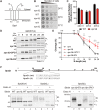
Signal sequence processing by SPase depends on the n region length and h region hydrophobicity of SSs. (A) Schematic of N#CPYt constructs. Left: blue lines indicate N-terminal extensions derived from the N terminus of the yeast membrane protein Dap2 (N-Dap2), and the black line indicates the yeast vacuole protein CPY. Numbers indicate the extended amino acids. N-linked glycosylation sites are shown as ‘Y’. HA, hemagglutinin tag. Right: diagram showing possible forms of N#CPYt variants in the ER. (B) Yeast transformants carrying the indicated N#CPYt(h) constructs were radiolabeled for 5 min at 30°C, immunoprecipitated using an anti-HA antibody, subjected to SDS–PAGE and analyzed by autoradiography. Endo H treatment was performed prior to SDS–PAGE. FL, full length; C, cleaved; gly., glycosylated species; degly., deglycosylated species. (C) Left: spc3-4 cells expressing N16CPYt(h) were incubated and radiolabeled for 5 min at the indicated temperatures. Right: the indicated N#CPYt(h) variants in the WT or spc3-4 strain were analyzed as in B, except that N#CPYt(h) variants in the spc3-4 strain were incubated at 37°C for 30 min prior to radiolabeling and were radiolabeled at 37°C. All the samples were treated with Endo H prior to SDS–PAGE. (D) The indicated CPY variants and Dap2 were subjected to carbonate extraction, and the resulting protein samples were detected by western blotting using an anti-HA antibody (Sup, supernatant). (E) Hydrophobicities of the N#CPYt variant SSs were predicted using the ΔG predictor (ΔGapp in kcal/mol; http://dgpred.cbr.su.se/index.php?p=home). Amino acids shown in bold indicate the modified residues from the SS of WT CPY. (F) The relative amounts of SPase-processed species over glycosylated products for each CPY variant were measured and plotted (as percentage cleavage). The x-axis indicates the number of amino acids (aa) preceding the SS (N-length). At least three independent experiments were carried out. Data are presented as mean±s.d. Data in B are representative of three independent experiments. Data in C and D are representative of two independent experiments.
Fig. 2.
Fig. 2.
Cleavage of internal SSs is enhanced in the absence of Spc1. (A) Schematics of the membrane topology of Spc1. Numbers indicate amino acid positions and highlight the ends of two TM domains of Spc1. (B) WT and spc1Δ cells were serially diluted from 0.2 OD600 cells and grown on YPD medium for 1 day at the indicated temperatures. Images are representative of three independent experiments. (C) The abundance of other SPC subunits, Sec11, Spc3 and Spc2, in the WT and spc1Δ strains was assessed using mass spectrometry. The mean±s.d. of three repeats is shown. (D) N#CPYt(h) variants in the WT, spc1Δ, spc1Δ+SPC1 and spc1Δ+EV strains were assessed as in Fig. 1B. All the samples were treated with Endo H prior to SDS–PAGE. (E) Percentage cleavage of N#CPYt(h) variants in the indicated strains was analyzed. At least three independent experiments were carried out. Data are presented as mean±s.d. P-values between WT and spc1Δ and between spc1Δ+EV and spc1Δ+SPC1 were calculated by multiple two-tailed, unpaired Student's t-tests and are shown in black and gray colors, respectively (n.s., P>0.05; **P≤0.01; ****P≤0.0001). (F) Top: schematic of Sps2t variants. Numbers indicate amino acid positions, glycosylation sites are shown as ‘Y’. Leucine mutation is shown in bold. Bottom: processing of Sps2t variants in WT, spc1Δ, spc1Δ+SPC1 and spc1Δ+EV strains. Experimental procedures were carried out as in Fig. 1B. Data are representative of three independent experiments. aa, amino acids; A.U., arbitrary units; C, cleaved; FL, full length; gly., glycosylated species.
Fig. 3.
Fig. 3.
Recognition and usage of the SS cleavage site by SPase is unchanged in the spc1Δ strain. (A) Two cleavage sites are present in the SS of N#CPYt(h): cleavage site 1 and cleavage site 2 are indicated as downward and upward arrows, respectively. (B) The indicated cleavage site mutants of N#CPYt(h) variants in the WT or spc3-4 strain were radiolabeled for 5 min at 30°C (37°C for spc3-4), immunoprecipitated using anti-HA antibodies, subjected to SDS–PAGE and Endo H treatment, and analyzed by autoradiography. (C) Percentage cleavage of the cleavage site mutants in B was analyzed as in Fig. 1F and compared. At least three independent experiments were carried out. Data are presented as mean±s.d. (D) Percentage cleavage of N#CPYt(h) variants with Q−3 or P+1′ mutations in the WT or spc1Δ strain is compared. At least three independent experiments were carried out. Data are presented as mean±s.d. (E) The indicated N#CPYt(h) variants lacking canonical cleavage sites in the WT or spc1Δ strain were radiolabeled and assayed as described in B. Data are representative of two independent experiments. aa, amino acids; C, cleaved; FL, full length.
Fig. 4.
Fig. 4.
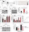
SPase-mediated processing of signal-anchored proteins is enhanced in the spc1Δ strain. (A) Schematic of LepCCt. The TM domain is colored black, and an N-linked glycosylation site is indicated as ‘Y’. A red arrowhead points to the site of cleavage by SPase. Flanking and TM sequences including the cleavage site (downwards arrow) are shown for three LepCCt variants. Leucine repeats are underlined. (B) The indicated LepCCt variants in WT cells were radiolabeled for 5 min at 30°C and subjected to immunoprecipitation for SDS–PAGE and autoradiography. Protein samples were treated with or without Endo H prior to SDS–PAGE. (C) The LepCCt(14L) construct in the WT or spc3-4 strain was radiolabeled and analyzed as in B. P+1 in LepCCt(14L) indicates proline substitution in the +1 position relative to the cleavage site. (D) LepCCt(17L) in the WT or spc1Δ strain was radiolabeled for 5 min and chased for the indicated time points at 30°C, then immunoprecipitated with anti-HA antibodies, subjected to SDS–PAGE and analyzed by autoradiography. Data in B are representative of three independent experiments. Data in C and D are representative of two independent experiments. C, cleaved species; FL, full length.
Fig. 5.
Fig. 5.
SPase-mediated processing of double-spanning membrane proteins is modulated by Spc1. (A) Schematics of LepH2. The second hydrophobic segment (H) of varying hydrophobicity is colored black. N-linked glycosylation sites are indicated as ‘Y’, and a red arrowhead points to the site of cleavage by SPase. 1G, singly glycosylated form; 2G, doubly glycosylated form. Amino acid sequences of the H segment are shown and underlined, with N- and C-terminal flanking residues. Leucine residues are highlighted. (B) LepH2(3L) in the WT or spc3-4 strain was radiolabeled for 5 min at 30°C. WT samples were treated with Endo H prior to SDS–PAGE. 0G, nonglycosylated form; 2G, doubly glycosylated form; C, cleaved form after membrane insertion. (C) LepH2(3L) in the WT+EV, spc1Δ+EV or spc1Δ+SPC1 strain was radiolabeled for 5 min at 30°C and analyzed. Left: autoradiogram of a representative blot is shown. Right: cleavage of the LepH2 H segments was calculated as a percentage of the cleaved band intensity over the sum of the 2G and cleaved band intensities from three independent experimental measurements. (D) Percentage membrane insertion of the H segment in LepH2 variants was measured as 2G/(total−0G)×100. (E) Left: percentage cleavage of the LepH2 H segments was calculated as in C. Right: relative cleavage of the LepH2 H segments in spc1Δ cells compared to that in WT cells was calculated as the ratio of percentage cleavage in spc1Δ cells and WT cells, and is plotted for each LepH2 variant. (F) LepH2(5L) in WT cells harboring control vector or Spc1 overexpression (OE) vector. Transformants were subjected to radiolabeling for 5 min at 30°C followed by a chase for the indicated time points. (G) Percentage cleavage of LepH2(5L) in F was calculated as in C. For all the experimental sets, at least three independent experiments were carried out, and data are presented as mean±s.d. *P≤0.05; **P≤0.01; ***P≤0.001; ****P≤0.0001; n.s., P>0.05 (two-tailed, unpaired Student's t-tests).
Fig. 6.
Fig. 6.
Overexpressed Spc1 interacts with model membrane proteins. Co-immunoprecipitation of overexpressed FLAG-tagged Spc1 with (A) N9CPYt and (B) LepCC variants. spc1Δ cells co-expressing Spc1–FLAG and the indicated HA-tagged substrates were subjected to crude membrane fractionation. Isolated membranes were solubilized with 1% Triton X-100 lysis buffer, followed by co-immunoprecipitation with anti-FLAG antibodies and visualization using SDS–PAGE and immunoblotting (IB) with the indicated antibodies. Representative blots from three experiments are shown. IP; immunoprecipitates; FL, full-length; C, cleaved. A red asterisk indicates full-length N9CPYt. Input lanes represent 2% of the total lysate.

References

    1. Almagro Armenteros, J. J., Tsirigos, K. D., Sønderby, C. K., Petersen, T. N., Winther, O., Brunak, S., von Heijne, G. and Nielsen, H. (2019). SignalP 5.0 improves signal peptide predictions using deep neural networks. Nat. Biotechnol. 37, 420-423. 10.1038/s41587-019-0036-z - DOI - PubMed
    1. Antonin, W., Meyer, H.-A. and Hartmann, E. (2000). Interactions between Spc2p and other components of the endoplasmic reticulum translocation sites of the yeast Saccharomyces cerevisiae. J. Biol. Chem. 275, 34068-34072. 10.1074/jbc.M006126200 - DOI - PubMed
    1. Barkocy-Gallagher, G. A. and Bassford, P. J.Jr. (1992). Synthesis of precursor maltose-binding protein with proline in the +1 position of the cleavage site interferes with the activity of Escherichia coli signal peptidase I in vivo. J. Biol. Chem. 267, 1231-1238. 10.1016/S0021-9258(18)48419-0 - DOI - PubMed
    1. Bird, P., Gething, M. J. and Sambrook, J. (1990). The functional efficiency of a mammalian signal peptide is directly related to its hydrophobicity. J. Biol. Chem. 265, 8420-8425. 10.1016/S0021-9258(19)38904-5 - DOI - PubMed
    1. Choo, K. H. and Ranganathan, S. (2008). Flanking signal and mature peptide residues influence signal peptide cleavage. BMC Bioinformatics 9 Suppl. 12, S15. 10.1186/1471-2105-9-S12-S15 - DOI - PMC - PubMed

Publication types

MeSH terms