Review
. 2021 Jul 7;13(7):e08253.
doi: 10.15252/emmm.201708253. Epub 2021 Jun 14.

Ang2 inhibitors and Tie2 activators: potential therapeutics in perioperative treatment of early stage cancer

Affiliations
Review

Ang2 inhibitors and Tie2 activators: potential therapeutics in perioperative treatment of early stage cancer

Kabir A Khan et al. EMBO Mol Med. .

Abstract

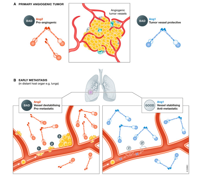
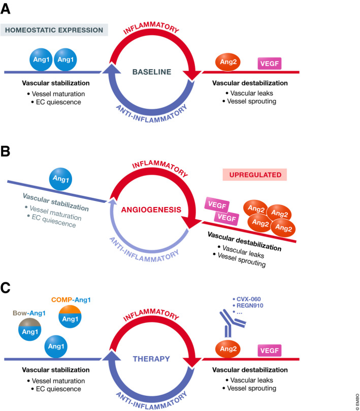
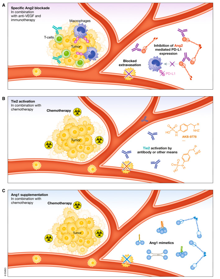
Anti-angiogenic drugs targeting the VEGF pathway are most effective in advanced metastatic disease settings of certain types of cancers, whereas they have been unsuccessful as adjuvant therapies of micrometastatic disease in numerous phase III trials involving early-stage (resectable) cancers. Newer investigational anti-angiogenic drugs have been designed to inhibit the Angiopoietin (Ang)-Tie pathway. Acting through Tie2 receptors, the Ang1 ligand is a gatekeeper of endothelial quiescence. Ang2 is a dynamically expressed pro-angiogenic destabilizer of endothelium, and its upregulation is associated with poor prognosis in cancer. Besides using Ang2 blockers as inhibitors of tumor angiogenesis, little attention has been paid to their use as stabilizers of blood vessels to suppress tumor cell extravasation and metastasis. In clinical trials, Ang2 blockers have shown limited efficacy in advanced metastatic disease settings. This review summarizes preclinical evidence suggesting the potential utility of Ang2 inhibitors or Tie2 activators as neoadjuvant or adjuvant therapies in the prevention or treatment of early-stage micrometastatic disease. We further discuss the rationale and potential of combining these strategies with immunotherapy, including immune checkpoint targeting antibodies.

Keywords: adjuvant; angiogenesis; immunotherapy; metastasis; neoadjuvant.


Conflict of interest statement

RSK is a member of the Scientific Advisory Board of Angiocrine Bioscience Inc., CSTS Healthcare, Novelty Nobility, Nonagen Therapeutics and OncoHost and a consultant to Novelty Nobility, Pharmabcine, and CSTS Healthcare. The other authors declare no conflicts of interest.

Figures

Figure 1
Figure 1. Schematic comparison of Ang1, Ang2, and engineered Ang1 variants/mimetics
(A, B) Structural domains of Angiopoietin‐1 (Ang1) and Angiopoietin‐2 (Ang2). FLD = fibrinogen‐like domain. CCD = coiled‐coil domain. SCD = superclustering domain. S = secretory signal. L = linker region. ECM = extracellular matrix. (C) Ang1 and Ang2 monomers form asymmetrical dimers where only one FLD is available to bind Tie2 (the light blue or light red FLD depicts the non‐binding monomer). (D) Ang1 dimers can form tetramers by oligomerization at the SCD, higher‐order oligomers can also be formed (not depicted here) (E) CMP‐Ang1 (modified coiled‐coil domain of cartilage matrix protein fused to Ang1 FLD) to form dimers. (F) MAT‐Ang1 (the coiled‐coil domain of matrillin fused to Ang1 FLD) form tetramers. (G) COMP‐Ang1 (cartilage oligomeric matrix protein fused to Ang1 FLD) form pentamers. (H) Ang1* contains N‐terminal SCD portions of Ang2 and also cysteine‐to‐serine mutations at residue 265 (or residue 245 in the mature protein without the signal peptide). C265S mutation depicted with *. (I) Bow‐Ang1 consists of four Ang1 FLDs fused to a dimerized Fc domain.
Figure 2
Figure 2. Schematic of Tie2 receptor domains, clustering, and activation
(A) Structural domains of monomeric Tie receptors. Angiopoietins bind the 2nd Ig domain (Ig2) of Tie2 (blue) but not of Tie1 (green). (B) In the absence of ligands, Tie2 can form inactive dimers through the membrane‐proximal Fn3 fibronectin type III domain (Leppänen et al, 2017; Moore et al, 2017). (C) Ang2 exists more commonly in lower‐order oligomeric forms, such as a dimer which does not activate Tie2, and rarely exists in tetramer and higher‐order forms, which can potentially explain its partial agonist activity. (D) Ang1 is the canonical full agonist of Tie2 that predominates in higher‐order forms, such as a tetramer, in which the two FLD’s available for Tie2 binding has been predicted to simultaneously engage two parallel Tie2 dimers within an arrayed complex as depicted (as opposed to the two arms of the same Tie2 dimer) (Leppänen et al, 2020). (E) Ang2 competes for the same binding site on Tie2 as Ang1 explaining its antagonistic effect on Ang1‐induced Tie2 phosphorylation. (F) Ang2‐binding Tie2‐activating Antibody (ABTAA) couples together two Ang2 dimers to enable tetrameric engagement and activation of Tie2. (F, G, and H) Tie2 clustering by engineered Ang1 variants/mimetics, Bow‐Ang1‐COMP‐Ang1, and CMP‐Ang1 are confirmed potent agonists of Tie2. As Bow‐Ang1 and COMP‐Ang1 do not contain the native coiled‐coil domains of Ang1, each FLD within the molecule is likely able to bind to a Tie2 ligand‐binding domain. (I) The small‐molecule inhibitor AKB‐9778 inhibits VE‐PTP, a phosphatase of Tie2, hence preserving Tie2 phosphorylation.
Figure 3
Figure 3. Differential roles of Ang1 and Ang2 in established growing tumors vs. metastatic dissemination
(A) At the site of established growing tumors, Ang2 acts as an angiogenesis initiator, while exogenous Ang1 diminishes the anti‐tumor efficacy of anti‐angiogenic anti‐VEGF and anti‐Ang2 therapies (Falcón et al, 2009; Huang et al, 2009; Coxon et al, 2010; Daly et al, 2013). (B) At the destination sites of metastasis, Ang2 de‐stabilizes the "normal" host blood vessels to facilitate various steps in the metastatic cascade (Talmadge & Fidler, 2010). (1) Tumor cell adhesion to capillary wall (Kim et al, 2001); (2) tumor cell extravasation (Gavard et al, 2008); and (3) tumor cell co‐option of existing host vessels (Holash et al, 1999). We thus hypothesize that Ang1, a vascular stabilizing factor, may inhibit these early metastatic events, i.e., in the lung: (1') inhibition of cancer cell adhesion and arrest (Michael et al, 2017); and (2') inhibition of extravasation due to stabilized vessels (Wu et al, 2015).
Figure 4
Figure 4. Targeting the Angiopoietins in Cancer Therapy
(A) Ang1 is constitutively expressed in low levels in healthy adults and maintains endothelial quiescence (Pfaff et al, 2006). (B) In many cancer types, overexpression of Ang2 and increased Ang2/Ang1 ratios correlate with more advanced disease stages and worse prognosis (Huang et al, 2010). (C) There are Ang2‐neutralizing agents already in clinical development as oncologic drugs, as well as preclinical investigatory agents activating Tie2.
Figure 5
Figure 5. Potential strategies for use of Ang/Tie targeting agents in combination with other therapies for inhibiting and/or treating early‐stage metastatic disease in, e.g., the lung
Tumor cells represent a micrometastatic lesion in a distant organ such as the lung. (A) Specific Ang2 blockade, along with its anti‐metastasis and vascular effects, blocks Ang2‐mediated upregulation of PD‐L1 on M2 macrophages. Ang2 blockade has the potential to reduce the immunosuppressive effects of PD‐L1 in the tumor microenvironment, which could be an ideal combination for immunotherapies such as agonist CD40 antibodies or PD‐1 blocking antibodies (green). Ang2 blockade also strengthens endothelial cell‐endothelial cell junctions inhibiting extravasation of tumor cells, along with a multitude of other effects described in the main text. Combining with VEGF blockade would also likely enhance the effects on the vasculature and relieving of immunosuppression. (B) Tie2 activation, whether induced by antibody drugs (AB‐Tie1‐39, ABTAA) or maintained by a small‐molecule inhibitor (AKB‐9778), potentially modulates organ vessels at possible sites of metastasis to reduce tumor cell extravasation and vessel co‐option. (C) Ang1 supplementation, in addition to activating Tie2, may additionally involve crosstalk with integrins to modulate vessels at metastatic sites and inhibit tumor cell extravasation. Moreover, both Ang1 supplementation and Tie2 activation may enhance the intratumoral delivery of chemotherapy, could be partnered with immunotherapy, and treat already seeded micrometastases, although this has not been described experimentally.

References

    1. Abdul Pari AA, Singhal M, Hübers C, Mogler C, Schieb B, Gampp A, Gengenbacher N, Reynolds LE, Terhardt D, Géraud C et al (2020) Tumor cell‐derived angiopoietin‐2 promotes metastasis in melanoma. Cancer Res 80: 2586–2598 - PMC - PubMed
    1. Adler AP, Daly C, Parveen AA, Nevins T, Shan J, Fairhurst J, Huang T, Martin J, Papadopoulos N, Yancopoulos GD et al (2014) Abstract 4492: Blockade of angiopoietin‐2 or Tie2 is equally effective at inhibiting tumor growth and reducing tumor vessel density in most human tumor xenograft models. Cancer Res 74: 4492
    1. Allegra CJ, Yothers G, O’Connell MJ, Sharif S, Petrelli NJ, Lopa SH, Wolmark N (2013) Bevacizumab in stage II‐III colon cancer: 5‐year update of the National Surgical Adjuvant Breast and Bowel Project C‐08 trial. J Clin Oncol 31: 359–364 - PMC - PubMed
    1. Allen E, Jabouille A, Rivera LB (2017) Combined antiangiogenic and anti–PD‐L1 therapy stimulates tumor immunity through HEV formation. Sci Transl Med 9: eaak9679 - PMC - PubMed
    1. Al Wadi K, Ghatage P (2016) Efficacy of trebananib (AMG 386) in treating epithelial ovarian cancer. Expert Opin Pharmacother 17: 853–860 - PubMed

Publication types

Grants and funding