. 2021 Jun 14;17(6):e1009068.
doi: 10.1371/journal.pcbi.1009068. eCollection 2021 Jun.

Free energy landscape of RNA binding dynamics in start codon recognition by eukaryotic ribosomal pre-initiation complex

Affiliations

Free energy landscape of RNA binding dynamics in start codon recognition by eukaryotic ribosomal pre-initiation complex

Takeru Kameda et al. PLoS Comput Biol. .

Abstract

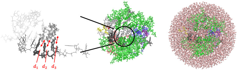
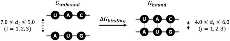
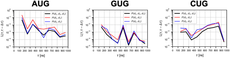
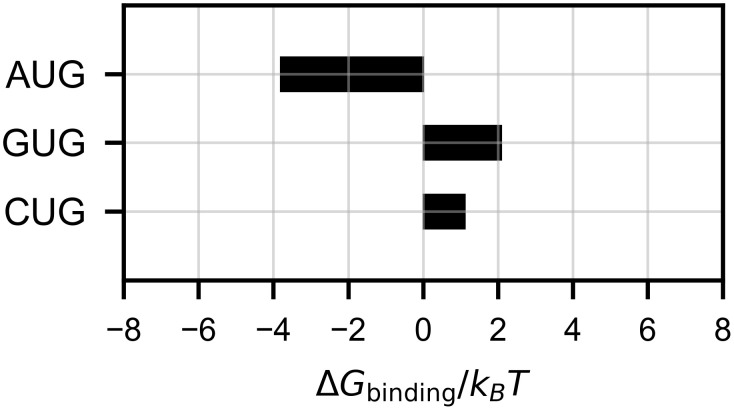
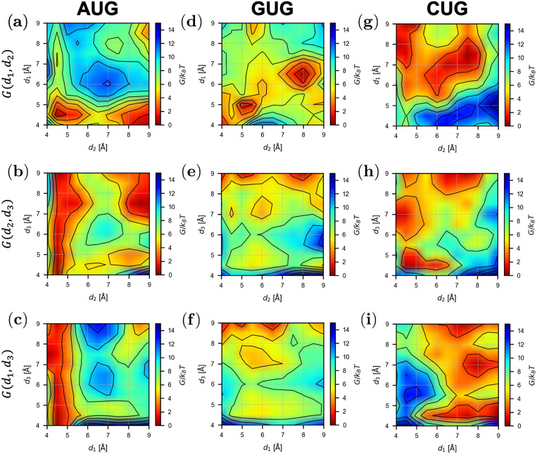
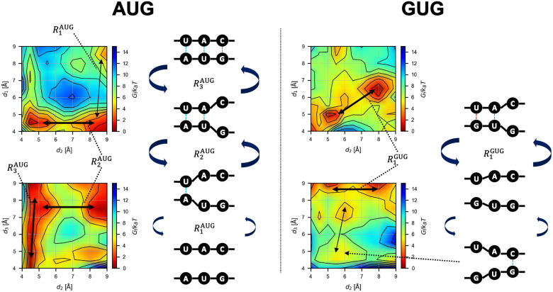
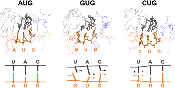
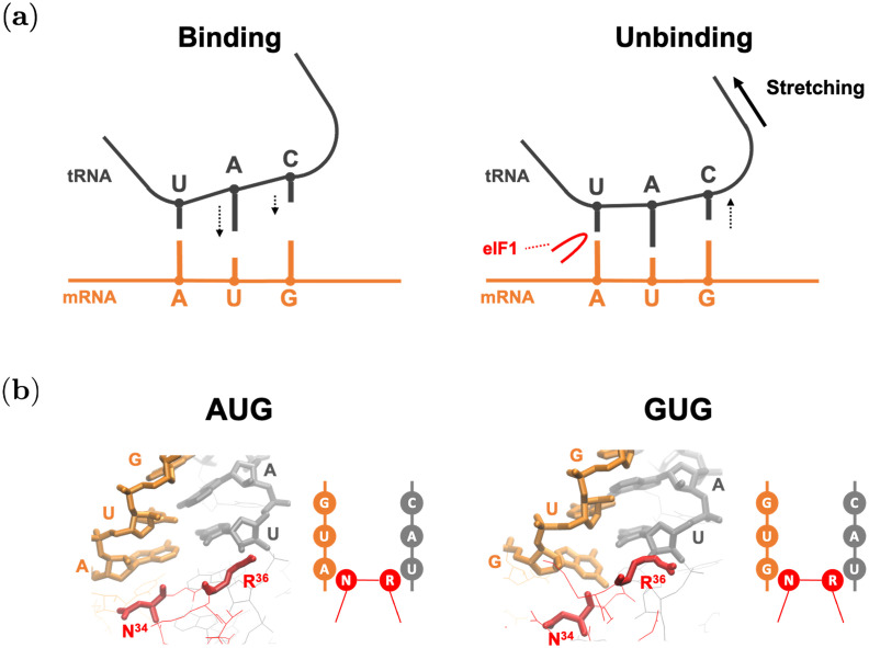
Specific interaction between the start codon, 5'-AUG-3', and the anticodon, 5'-CAU-3', ensures accurate initiation of translation. Recent studies show that several near-cognate start codons (e.g. GUG and CUG) can play a role in initiating translation in eukaryotes. However, the mechanism allowing initiation through mismatched base-pairs at the ribosomal decoding site is still unclear at an atomic level. In this work, we propose an extended simulation-based method to evaluate free energy profiles, through computing the distance between each base-pair of the triplet interactions involved in recognition of start codons in eukaryotic translation pre-initiation complex. Our method provides not only the free energy penalty for mismatched start codons relative to the AUG start codon, but also the preferred pathways of transitions between bound and unbound states, which has not been described by previous studies. To verify the method, the binding dynamics of cognate (AUG) and near-cognate start codons (CUG and GUG) were simulated. Evaluated free energy profiles agree with experimentally observed changes in initiation frequencies from respective codons. This work proposes for the first time how a G:U mismatch at the first position of codon (GUG)-anticodon base-pairs destabilizes the accommodation in the initiating eukaryotic ribosome and how initiation at a CUG codon is nearly as strong as, or sometimes stronger than, that at a GUG codon. Our method is expected to be applied to study the affinity changes for various mismatched base-pairs.


Conflict of interest statement

The authors have declared that no competing interests exist.

Figures

Fig 1
Fig 1. PIC structure models and reaction coordinates in this study.
(Left) mRNA and tRNA around the codon-anticodon base-pairs (Table 1) are shown in black and silver, respectively. Distances d1, d2 and d3 used as reaction coordinates are indicated. (Center) rRNA segments (split) are shown in green, and protein components are shown in other colors. (Right) Water molecules are shown in pink, which enclose the system and form a sphere.
Fig 2
Fig 2. Binding free energy.
Schematic representation of Gbound, Gunbound, and ΔGbinding (Eq 3).
Fig 3
Fig 3. Convergence of free energy profiles with time evolution.
The L(τ, τ − Δτ) of P(d1, d2, d3), P(d1, d2), and P(d2, d3) are shown (see Eq 5; Δτ = 100 ns). The data points are plotted between the two consecutive time points (τ − Δτ) and τ.
Fig 4
Fig 4. Estimated binding free energy.
ΔGbinding scores (Eq 3; see also Fig 2) are shown. The scores were obtained from P(d1, d2, d3) averaged over five simulation trials for each model.
Fig 5
Fig 5. Projected free energy profiles.
Profiles of G(d1, d2), G(d2, d3), and G(d1, d3) obtained from Eq 4 for each model is shown by contour plots.
Fig 6
Fig 6. Schematics of the base-pair binding dynamics.
Conformational changes inferred from the free energy landscape (Fig 5). The transition path Rn (• is AUG or GUG) is shown by black arrows (thick arrow: frequent transition; thin arrow: less frequent transition). Blue and red dotted lines indicate Watson-Crick or wobble base-pairing, respectively. In left, AUG pairing is described as being stabilized by the first position. Subsequently, the binding propagates into the second and the third position. In right, GUG pairing is described as bistable states; the base-pairing between GU and the corresponding bases in the anticodon, and the base pairing between the 3rd G and the C residue of the anticodon. Thus, a complete base-triplet interaction is unstable.
Fig 7
Fig 7. Typical bound structures.
Averaged structures corresponding to reaction coordinate (d1˜,d2˜,d3˜)=(4.5,4.5,4.5) (see Eq 6). In the structures on top, nucleotides of the codon (orange) and anticodon (gray) are drawn by thick lines. Red and blue lines are parts of eIF1 and eIF1A, respectively. Note that the structures of GUG and CUG pairing (middle and right) are much less stable than that of AUG pairing (left) (see Fig 4). The schematics on the bottom describe the direction of each base relative to the paired base.
Fig 8
Fig 8. Binding mechanism conjectured by this study.
mRNA and tRNA are drawn by orange and gray, respectively. Red lines show parts of eIF1. (a) Schematics of binding and unbinding dynamics of AUG pairing in the P-site. (Left) AUG binding is facilitated by the stabilization of the first base-pair, followed by base-pairing of the 2nd and 3rd positions (downward arrows). (Right) AUG unbinding appears to be facilitated by dissociation of the 3rd position (upward arrow) through stretching (thick arrow) imposed by the open (POUT) conformation of the PIC [11]. Here, eIF1 β-hairpin loop (red) is proposed to prevent dissociation of the base-pairing at the 1st position [35]. (b) Averaged structures corresponding to reaction coordinate (d1˜,d2˜,d3˜)=(4.5,4.5,4.5) (see Eq 6), and their schematic representations. Nucleotides of the codon and anticodon, and N34 (Asn-34) and R36 (Arg-36) are drawn by thick lines.

References

    1. Asano K. Translational control. In: Encyclopedia of Systems Biology. Springer; New York; 2013. p. 2278–2282.
    1. Asano K, Ito K. Translation elongation. In: Encyclopedia of Systems Biology. Springer; New York; 2013. p. 2259–2263.
    1. Hinnebusch AG, Dever TE, Asano K. Mechanism of translation initiation in the yeast Saccharomyces cerevisiae. Cold Spring Harbor Monograph Series. 2007;48:225–268.
    1. Sonenberg N, Hinnebusch AG. Regulation of translation initiation in eukaryotes: mechanisms and biological targets. Cell. 2009;136(4):731–745. doi: 10.1016/j.cell.2009.01.042 - DOI - PMC - PubMed
    1. Asano K, Clayton J, Shalev A, Hinnebusch AG. A multifactor complex of eukaryotic initiation factors, eIF1, eIF2, eIF3, eIF5, and initiator tRNAMet is an important translation initiation intermediate in vivo. Genes Dev. 2000;14(19):2534–2546. doi: 10.1101/gad.831800 - DOI - PMC - PubMed

Publication types

MeSH terms