. 2021 Jun 14;17(6):e1009656.
doi: 10.1371/journal.ppat.1009656. eCollection 2021 Jun.

Conservative production of galactosaminogalactan in Metarhizium is responsible for appressorium mucilage production and topical infection of insect hosts

Affiliations

Conservative production of galactosaminogalactan in Metarhizium is responsible for appressorium mucilage production and topical infection of insect hosts

Lijuan Mei et al. PLoS Pathog. .

Abstract

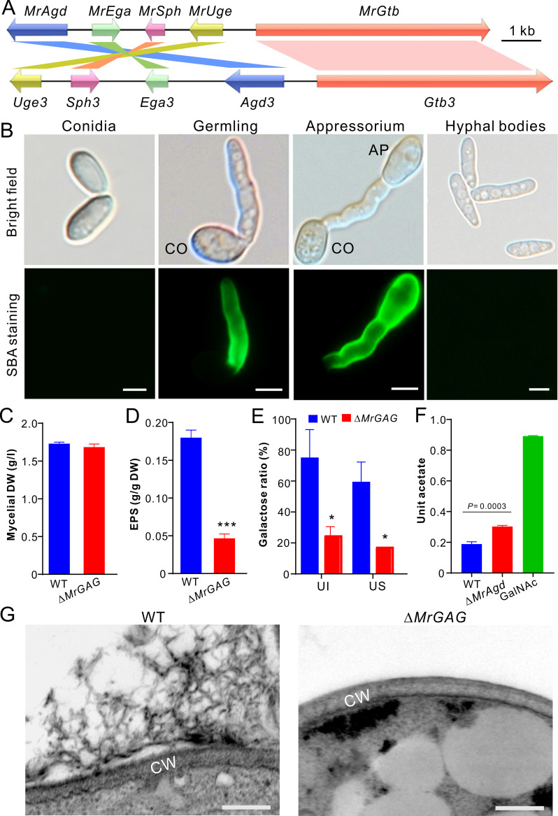
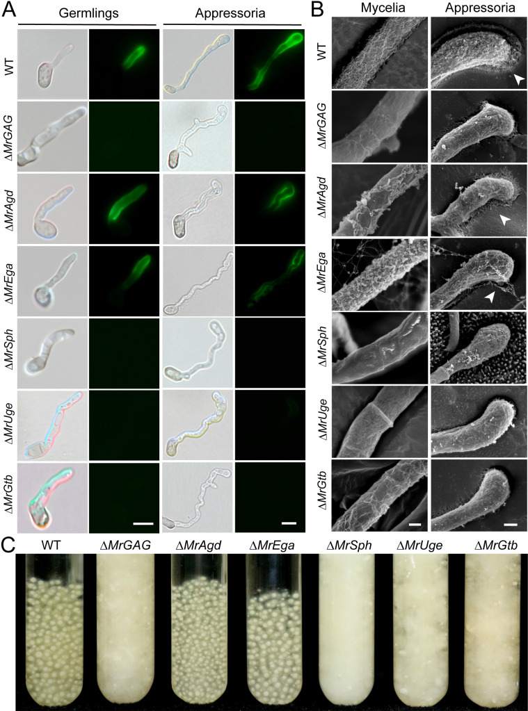
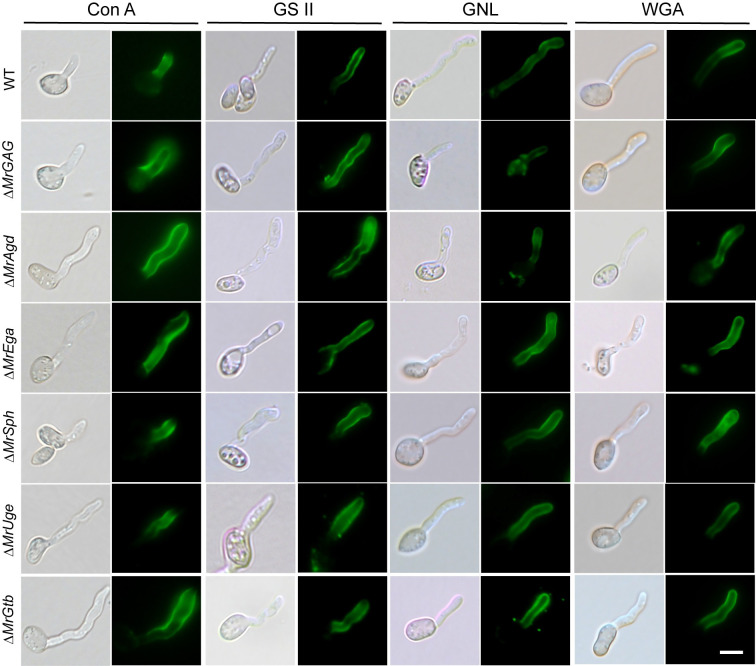
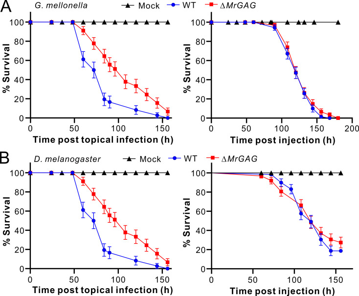
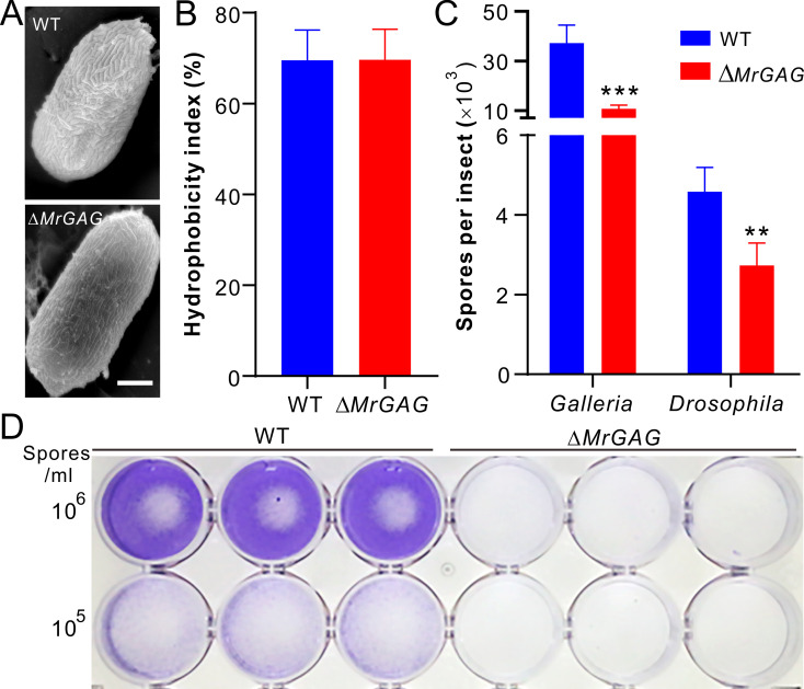
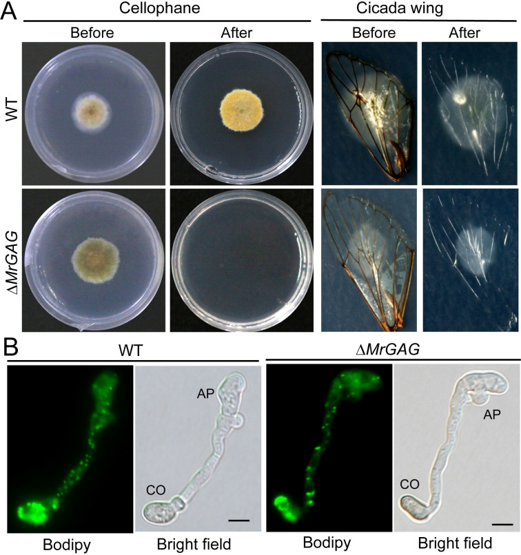
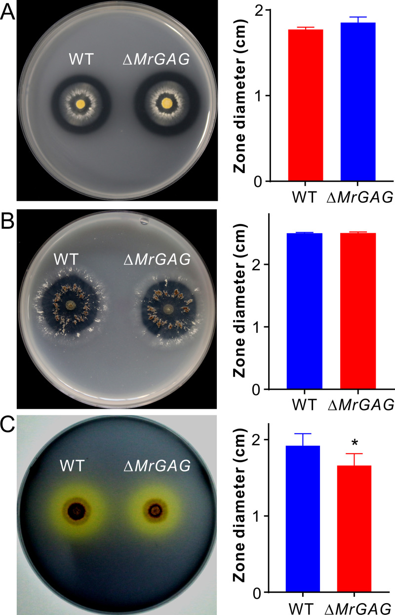
The exopolysaccharide galactosaminogalactan (GAG) has been well characterized in Aspergilli, especially the human pathogen Aspergillus fumigatus. It has been found that a five-gene cluster is responsible for GAG biosynthesis in Aspergilli to mediate fungal adherence, biofilm formation, immunosuppression or induction of host immune defences. Herein, we report the presence of the conserved GAG biosynthetic gene cluster in the insect pathogenic fungus Metarhizium robertsii to mediate either similar or unique biological functions. Deletion of the gene cluster disabled fungal ability to produce GAG on germ tubes, mycelia and appressoria. Relative to the wild type strain, null mutant was impaired in topical infection but not injection of insect hosts. We found that GAG production by Metarhizium is partially acetylated and could mediate fungal adherence to hydrophobic insect cuticles, biofilm formation, and penetration of insect cuticles. In particular, it was first confirmed that this exopolymer is responsible for the formation of appressorium mucilage, the essential extracellular matrix formed along with the infection structure differentiation to mediate cell attachment and expression of cuticle degrading enzymes. In contrast to its production during A. fumigatus invasive growth, GAG is not produced on the Metarhizium cells harvested from insect hemocoels; however, the polymer can glue germ tubes into aggregates to form mycelium pellets in liquid culture. The results of this study unravel the biosynthesis and unique function of GAG in a fungal system apart from the aspergilli species.


Conflict of interest statement

The authors have declared that no competing interests exist.

Figures

Fig 1
Fig 1. Biosynthesis of GAG in M. robertsii.
(A) Mesosyntenic relationship of the GAG biosynthetic gene clusters between M. robertsii and A. fumigatus. (B) Variation of GAG production by different type of cells. CO, conidium; AP, appressorium. Bar, 5 μm. (C) No difference of mycelium biomass production between WT and ΔMrGAG. The fungi were grown in SDB for three days and the mycelia were harvested, dried and weighted. DW, dry weight. (D) Comparison of the crude exopolysaccharide (EPS) production between WT and ΔMrGAG. Fungal samples were collected three days post inoculation in SDB. The urea-insoluble pellets were freeze-dried and compared. The significance of the two-tailed Student’s t-test difference is at: ***, P < 0.001. (E) Comparison of the galactose content hydrolyzed from the EPS produced by WT and ΔMrGAG. Fungal samples were harvested three days post inoculation in SDB. UI, urea insoluble EPS sample; US, urea soluble EPS sample. (F) Comparative quantification analysis of GAG acetylation. The EPS harvested from the WT and ΔMrAgd culture filtrates were hydrolyzed prior to the analysis of both reducing sugar and acetate. Unit acetate is referred to the acetate content out of the reducing sugar within each hydrolyzed sample. The standard GalNAc was hydrolyzed and used as a control. (G) Mycelial surface structure differences. The WT and ΔMrGAG mycelia were harvested from the day 3 SDB cultures for TEM analysis. Bar, 100 nm. CW, cell wall.
Fig 2
Fig 2. Microscopic and liquid-culture phenotyping.
(A) Fluorescent staining of the WT and mutant cells. Fungal cells were germinated in SDB for 12 hrs or induced for appressorium production in a MM medium for 18 hrs prior to the staining with SBA. Bar, 10 μm. (B) SEM observation of fungal mycelium and appressorium cells. The mycelia of the WT and mutants harvested from the day 3 SDB medium were used for SEM analysis. The appressoria of the WT and mutants were induced on the mealworm front wings. Mucilage matrixes produced by the WT, ΔMrAgd and ΔMrEga appressoria are arrowed. Bar, 200 nm. (C) Phenotyping of the liquid cultures of the WT and different mutants. Relative to the WT, production or non-production of mycelial pellets was observed for different mutants after the growth in SDB for three days.
Fig 3
Fig 3. Lectin staining of the WT and mutant germlings for different cell wall constituents.
The WT and mutant spores were germinated in SDB and then stained with different fluorescent lectins: ConA, Concanavalin A for detecting α-mannopyranosyl and α-glucopyranosyl residues; GSII for detecting α- or β-linked N-acetyl-D-glucosamine); GNL, Galanthus nivalis lectin for detecting α-1,3-mannose; WGA, wheat germ agglutinin for selective binding N-acetyl-D-glucosamine and N-acetylneuraminic acid residues.
Fig 4
Fig 4. Insect survivals after infections by WT and ΔMrGAG.
(A) Topical infection (left panel) and injection (right panel) assays against the last instar larvae of wax moth G. mellonella. (B) Topical infection (left panel) and injection (right panel) assays against the female adults of the fruit fly D. melanogaster. Insects treated with 0.05% Tween-20 were used as a mock control.
Fig 5
Fig 5. Conidial hydrophobicity and adhesion assays.
(A) SEM observation of the WT and mutant conidia. Bar, 2 μm. (B) No difference of the conidial spore hydrophobicity between WT and ΔMrGAG. (C) Variation of the spore adhesion abilities between WT and ΔMrGAG toward different insects. The spores were washed off and counted eight hours post treatment. The significance of the two-tailed Student’s t-test difference is at: ***, P < 0.001; **, P < 0.01. (D) Variation of the spore adhesion ability between WT and ΔMrGAG toward hydrophobic surface. Different concentration of spore suspensions were inoculated into the 24-well plate for 24 hrs and then washed off with PBS buffer. The wells were stained with crystal violet before imaging.
Fig 6
Fig 6. Penetration and appressorium formation assays.
(A) Fungal penetration assays. Both the WT and ΔMrGAG were inoculated on cellophane membrane for three days or cicada wings for 40 hrs. The membrane and insect wings were then carefully removed with fungal cultures, and the plates were kept for incubation for another one week before photographing. (B) Appressorium formation and lipid-droplet utilization by WT and ΔMrGAG. Appressorium were induced on hydrophobic surfaces for 18 hrs and stained with the fluorescent dye Bodipy. CO, conidium; AP, appressorium. Bar, 5 μm.
Fig 7
Fig 7. Comparative enzyme hydrolytic activity assays between WT and ΔMrGAG.
(A) Non-significant variation between strains in digestion of casein seven days post inoculation. (B) Non-significant variation between strains in digestion of chitin seven days post inoculation. (C) Variation between strains in digestion of olive oil five days post inoculation. The right part of each panel shows the data of the hydrolytic zone diameters formed by WT and ΔMrGAG. The significance of the two-tailed Student’s t-test difference is at: *, P = 0.03.

References

    1. Latgé J-P, Beauvais A, Chamilos G. The cell wall of the human fungal pathogen Aspergillus fumigatus: biosynthesis, organization, immune response, and virulence. Annu Rev Microbiol. 2017; 71:99–116. doi: 10.1146/annurev-micro-030117-020406 . - DOI - PubMed
    1. Brown L, Wolf JM, Prados-Rosales R, Casadevall A. Through the wall: extracellular vesicles in Gram-positive bacteria, mycobacteria and fungi. Nat Rev Microbiol. 2015; 13:620–630. doi: 10.1038/nrmicro3480 - DOI - PMC - PubMed
    1. Boutrot F, Zipfel C. Function, Discovery, and Exploitation of Plant Pattern Recognition Receptors for Broad-Spectrum Disease Resistance. Annu Rev Phytopathol. 2017; 55:257–286. doi: 10.1146/annurev-phyto-080614-120106 . - DOI - PubMed
    1. Lu Y, Su F, Li Q, Zhang J, Li Y, Tang T, et al. Pattern recognition receptors in Drosophila immune responses. Dev Comp Immunol. 2019; 102:103468. doi: 10.1016/j.dci.2019.103468 . - DOI - PubMed
    1. Mahapatra S, Banerjee D. Fungal exopolysaccharide: production, composition and applications. Microbiol Insights. 2013; 6:1–16. doi: 10.4137/MBI.S10957 . - DOI - PMC - PubMed

Publication types

MeSH terms

Supplementary concepts