. 2021 Oct;100(4):915-927.
doi: 10.1016/j.kint.2021.05.032. Epub 2021 Jun 11.

Adaptive lymphocyte profile analysis discriminates mild and severe forms of COVID-19 after solid organ transplantation

Affiliations

Adaptive lymphocyte profile analysis discriminates mild and severe forms of COVID-19 after solid organ transplantation

Arnaud Del Bello et al. Kidney Int. 2021 Oct.

Abstract

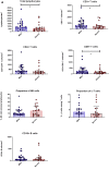
Solid organ transplant recipients are at high risk for the development of severe forms of COVID-19. However, the role of immunosuppression in the morbidity and mortality of the immune phenotype during COVID-19 in transplant recipients remains unknown. In this retrospective study, we compared peripheral blood T and B cell functional and surface markers, as well as serum antibody development during 29 cases of mild (World Health Organization 9-point Ordinal Scale (WOS) of 3-4) and 22 cases of severe COVID-19 (WOS 5-8) in solid organ transplant (72% kidney transplant) recipients hospitalized in our center. Patients who developed severe forms of COVID-19 presented significantly lower CD3+ (median 344/mm3 (inter quartile range 197; 564) vs. 643/mm3 (397; 1251)) and CD8+ T cell counts (124/mm3 (76; 229) vs. 240/mm3 (119; 435)). However, activated CD4+ T cells were significantly more frequent in severe forms (2.9% (1.37; 5.72) vs. 1.4% (0.68; 2.35)), counterbalanced by a significantly higher proportion of Tregs (3.9% (2.35; 5.87) vs. 2.7% (1.9; 3.45)). A marked decrease in the proportion of NK cells was noted only in severe forms. In the B cell compartment, transitional B cells were significantly lower in severe forms (1.2% (0.7; 4.2) vs. 3.6% (2.1; 6.2)). Nonetheless, a majority of transplant recipients developed antibodies against SARS-CoV-2 (77% and 83% in mild and severe forms, respectively). Thus, our data revealed immunological differences between mild and severe forms of COVID-19 in solid organ transplant recipients, similar to previous reports in the immunocompetent population.

Keywords: COVID-19; NK cells; exhaustion; lymphopenia; organ transplantation; regulatory T cells.


Figures

None
Graphical abstract
Figure 1
Figure 1
(a–e)Comparison of natural killer (NK), conventional and regulatory T (Treg)–, and B-cell compartments in mild and severe forms of coronavirus disease 2019. Severe forms were analyzed using the immunological sample taken at the closest time before the worse clinical situation. (a) Total lymphocyte count; CD3+, CD4+, and CD8+ T-cell count; and proportion of NK cells, δγ T cells, and CD19+ B cells. (b) T-cell compartment: CD4+ memory T-cell compartment, Tregs, activation senescence and exhaustion markers, and functional markers. CD8+ memory T-cell compartment, activation senescence and exhaustion markers, and functional markers. (c) Correlation between the percentage of Tregs and CD4+TIGIT+ or CD4+CD39+ cells, between the percentage of Tregs and CD8+PD-1+ or CD8+CD39+, and between the percentage of Tregs and CD4+TIGIT+ and CD4+perforin+/granzyme B+ (CD4+PRF+/GZM+) in mild and severe forms. The linear regression analysis was assessed using a Pearson correlation. (d) B-cell compartment: naive, transitional, activated, memory B cells and plasmablasts. Data are expressed as median with interquartile range. (e) Correlation between the percentage of CD27+ memory B cells and CD4+ effector memory (EM) T cells in mild and severe forms. The linear regression analysis was assessed using a Pearson correlation. ∗P < 0.05. CM, central memory; EMRA, effector memory re-expressing CD45RA; PD-1, programmed death receptor-1; TIGIT, T-cell immunoreceptor with Ig and ITIM domains.
Figure 1
Figure 1
(a–e)Comparison of natural killer (NK), conventional and regulatory T (Treg)–, and B-cell compartments in mild and severe forms of coronavirus disease 2019. Severe forms were analyzed using the immunological sample taken at the closest time before the worse clinical situation. (a) Total lymphocyte count; CD3+, CD4+, and CD8+ T-cell count; and proportion of NK cells, δγ T cells, and CD19+ B cells. (b) T-cell compartment: CD4+ memory T-cell compartment, Tregs, activation senescence and exhaustion markers, and functional markers. CD8+ memory T-cell compartment, activation senescence and exhaustion markers, and functional markers. (c) Correlation between the percentage of Tregs and CD4+TIGIT+ or CD4+CD39+ cells, between the percentage of Tregs and CD8+PD-1+ or CD8+CD39+, and between the percentage of Tregs and CD4+TIGIT+ and CD4+perforin+/granzyme B+ (CD4+PRF+/GZM+) in mild and severe forms. The linear regression analysis was assessed using a Pearson correlation. (d) B-cell compartment: naive, transitional, activated, memory B cells and plasmablasts. Data are expressed as median with interquartile range. (e) Correlation between the percentage of CD27+ memory B cells and CD4+ effector memory (EM) T cells in mild and severe forms. The linear regression analysis was assessed using a Pearson correlation. ∗P < 0.05. CM, central memory; EMRA, effector memory re-expressing CD45RA; PD-1, programmed death receptor-1; TIGIT, T-cell immunoreceptor with Ig and ITIM domains.
Figure 2
Figure 2
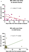
(a,b) Comparison of natural killer (NK), conventional T-, and B-cell compartments in an initially mild case that became severe after the first blood test and those that remained mild. (a) NK cell count over time in mild and severe forms. (b) CD3+, activated PD-1+CD4+, and CD39+CD4+ T cells, perforin+/granzyme B+CD4+ T cells, CD8+ T cells, and memory CD19+CD21low B cells. Data are expressed as mean with SEM. ∗P < 0.05. PD-1, programmed death receptor-1.
Figure 2
Figure 2
(a,b) Comparison of natural killer (NK), conventional T-, and B-cell compartments in an initially mild case that became severe after the first blood test and those that remained mild. (a) NK cell count over time in mild and severe forms. (b) CD3+, activated PD-1+CD4+, and CD39+CD4+ T cells, perforin+/granzyme B+CD4+ T cells, CD8+ T cells, and memory CD19+CD21low B cells. Data are expressed as mean with SEM. ∗P < 0.05. PD-1, programmed death receptor-1.

References

    1. Akalin E., Azzi Y., Bartash R. Covid-19 and kidney transplantation. N Engl J Med. 2020;382:2475–2477. - PMC - PubMed
    1. Nair V., Jandovitz N., Hirsch J.S. COVID-19 in kidney transplant recipients. Am J Transplant. 2020;20:1819–1825. - PMC - PubMed
    1. Wu C., Chen X., Cai Y. Risk factors associated with acute respiratory distress syndrome and death in patients with coronavirus disease 2019 pneumonia in Wuhan, China. JAMA Intern Med. 2020;180:934–943. - PMC - PubMed
    1. Huang C., Wang Y., Li X. Clinical features of patients infected with 2019 novel coronavirus in Wuhan, China. Lancet. 2020;395:497–506. - PMC - PubMed
    1. Hadjadj J., Yatim N., Barnabei L. Impaired type I interferon activity and inflammatory responses in severe COVID-19 patients. Science. 2020;369:718–724. - PMC - PubMed