. 2021 Oct 10:518:59-71.
doi: 10.1016/j.canlet.2021.06.007. Epub 2021 Jun 12.

Hedgehog transcriptional effector GLI mediates mTOR-Induced PD-L1 expression in gastric cancer organoids

Affiliations

Hedgehog transcriptional effector GLI mediates mTOR-Induced PD-L1 expression in gastric cancer organoids

Vivien Koh et al. Cancer Lett. .

Abstract

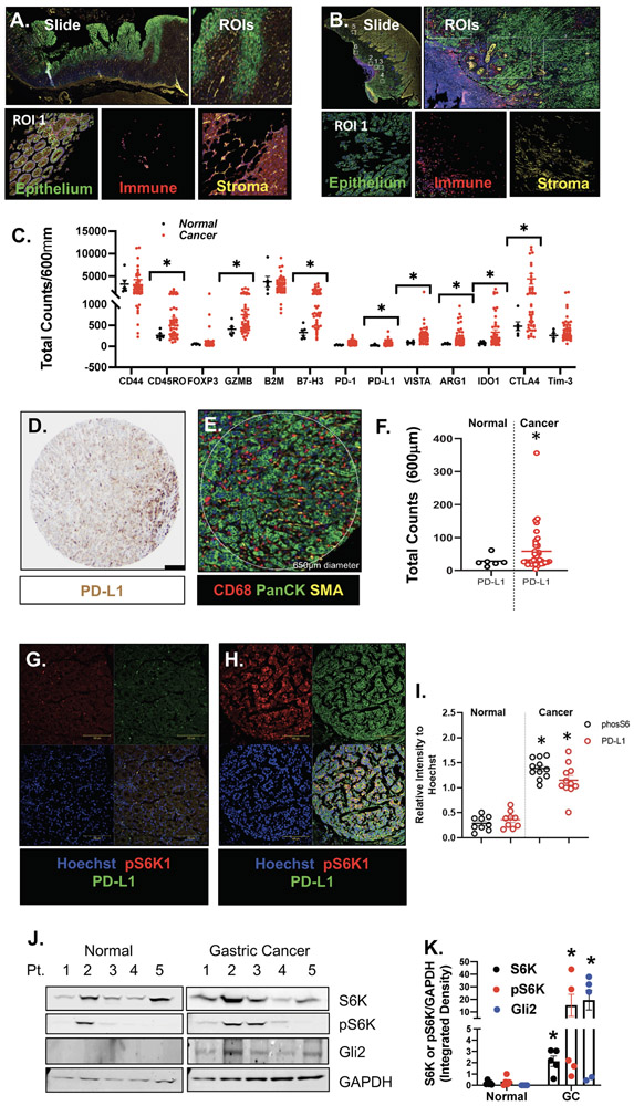
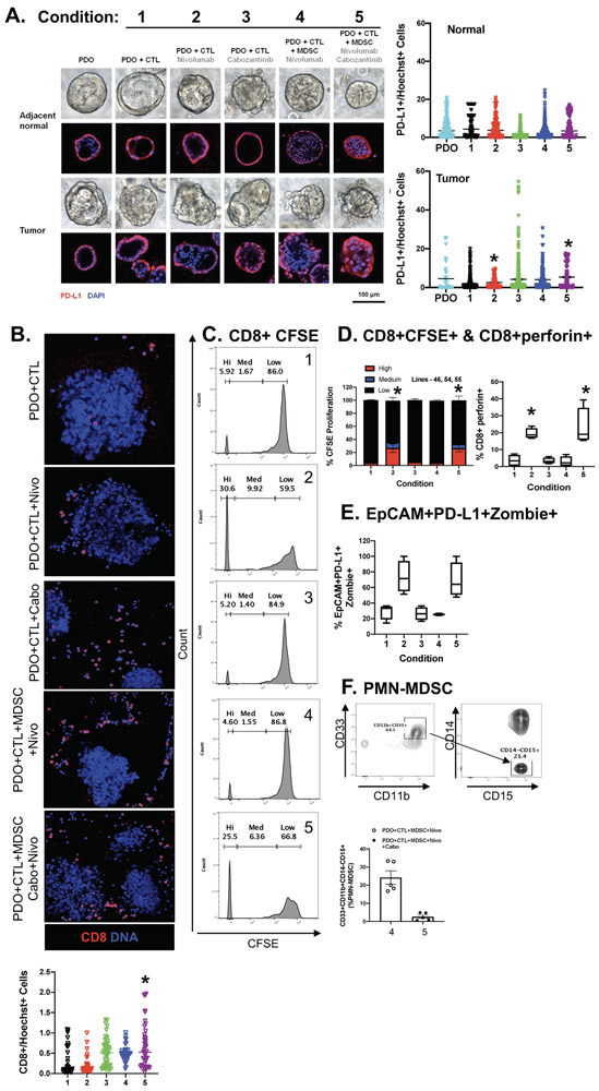
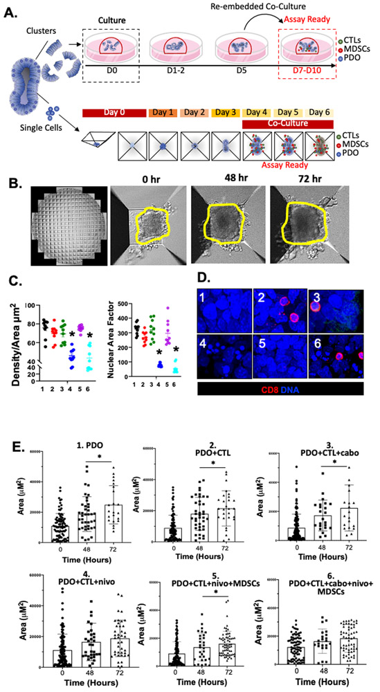
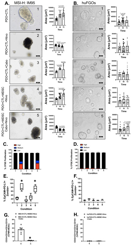
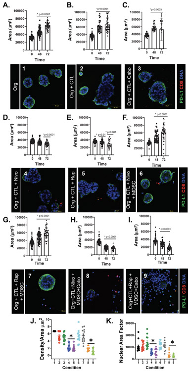
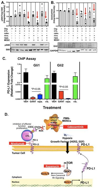
Tumors evade immune surveillance by expressing Programmed Death-Ligand 1 (PD-L1), subsequently inhibiting CD8+ cytotoxic T lymphocyte function. Response of gastric cancer to immunotherapy is relatively low. Our laboratory has reported that Helicobacter pylori-induced PD-L1 expression within the gastric epithelium is mediated by the Hedgehog (Hh) signaling pathway. The PI3K/AKT/mTOR pathway is activated in gastric cancer and may have immunomodulatory potential. We hypothesize that Hh signaling mediates mTOR-induced PD-L1 expression. Patient-derived organoids (PDOs) were generated from gastric biopsies and resected tumor tissues. Autologous organoid/immune cell co-cultures were used to study the immunosuppressive function of MDSCs. NanoString Digital Spatial Profiling (DSP) of immune-related protein markers using FFPE slide-mounted tissues from gastric cancer patients was performed. DSP analysis showed infiltration of immunosuppressive MDSCs expressing Arg1, CD66b, VISTA and IDO1 within cancer tissues. Orthotopic transplantation of patient derived organoids (PDOs) resulted in the engraftment of organoids and the development of histology similar to that observed in the patient's tumor tissue. PDO/immune cell co-cultures revealed that PD-L1-expressing organoids were unresponsive to nivolumab in vitro in the presence of PMN-MDSCs. Depletion of PMN-MDSCs within these co-cultures sensitized the organoids to anti-PD-1/PD-L1-induced cancer cell death. Rapamycin decreased phosphorylated S6K, Gli2 and PD-L1 expression in PDO/immune cell co-cultures. Transcriptional regulation of PD-L1 by GLI1 and GLI2 was blocked by rapamycin. In conclusion, the PDO/immune cell co-cultures may be used to study immunosuppressive MDSC function within the gastric tumor microenvironment. The mTOR signaling pathway mediates GLI-induced PD-L1 expression in gastric cancer.

Keywords: Cytotoxic T lymphocytes; Myeloid derived suppressor cells; Sonic hedgehog; Tumor microenvironment.


Figures

Figure 1:
Figure 1:. DSP analysis of FFPE tissues collected from normal and cancer gastric tissues.
Representative ROIs selected in (A) normal and (B) gastric cancer tissue. (C) Protein expression counts of immune (CD68+) cell markers in normal (black) and gastric cancer (red) tissues. (D) IHC staining showing expression of PD-L1 and (E) corresponding DSP ROI. (F) Protein expression counts of PD-L1 in PanCK+ normal (black) and gastric cancer (red) tissues. (G, H) Immunofluorescence using antibodies specific for the expression of PD-L1 (green) and phosphorylated S6K (pS6K1, red) in normal and cancer tissues. (I) Relative intensity of pS6K (black) and PD-L1 (red). (J) Western blots for the expression of S6K, pS6K, GLI2 and GAPDH in normal and gastric cancer patient tissues. (K) Densitometric analysis of Western blots. *p<0.05 compared to normal, n = 6 normal and n = 50 gastric cancer patients.
Figure 2:
Figure 2:. Effect of PMN-MDSCs in PD-L1+ PDO/immune cell co-cultures treated with nivolumab.
(A) Morphological changes in PDOs after 48 hours of in vitro treatment with vehicle (Control) alone, vehicle (Control) in co-cultures with CTLs (Condition 1), nivolumab (0.5 μg/ml; Condition 2) or Cabozantinib (10 μM; Condition 3) in co-cultures with CTLs, nivolumab (Condition 4) or a combination of nivolumab and Cabozantinib (Condition 5) in co-cultures with CTLs and MDSCs. (B) Images showing changes in organoid viability and CD8+ cell expression in organoid co-cultures. (C) Measurement of changes in CTL proliferation by CFSE, with quantification shown in (D). Changes in (D) perforin expression in CTLs, (E) percent of viable EpCAM+/PD-L1+ organoids and (F) PMN-MDSCs in response to treatments. *p<0.05 compared to condition 1, n = 4 organoid co-cultures derived from individual patients.
Figure 3:
Figure 3:. Morphological changes in PD-L1+ PDO/immune cell co-cultures in response to nivolumab.
(A) Schematic diagram showing the generation and timeline of organoid/immune cell co-culture using either Matrigel® dome or AggreWell™ microwell plates. (B) Representative images of morphological changes of PDO/immune cell co-cultures after 0, 48 and 72 hours of treatment in AggreWell™ microplates (yellow outlining indicates increased organoid area. (C) Changes in Density/Area (μm2) and nuclear area factor 72 hours after treatment in conditions 1 - 6. (D) Immunofluorescence showing expression of CD8+ cells (red) and nuclei stained by DAPI 72 hours after treatment in conditions 1 – 6. (E) Change in organoid Area ((μm2) in vehicle (condition 1) in co-cultures with CTLs (condition 2), cabozantinib (condition 3) or nivolumab (condition 4) in co-cultures with CTLs, nivolumab (condition 5) or a combination of nivolumab and cabozantinib (condition 6) in co-cultures with CTLs and MDSCs at 0, 48 and 72 hours. *p<0.05 compared to 0hr/vehicle treated for each condition besides condition 4 and 6 (ns= nonsignificant), n = 4 organoid co-cultures derived from individual patients.
Figure 4:
Figure 4:. Effect of PMN-MDSCs on CTL proliferation and cancer cell death in PD-L1+ PDO/immune cell co-cultures.
Changes in morphology and Area (μm2) of (A) MSI-H IM95 gastric cancer/immune cell co-cultures, and (B) huFGO/immune cell co-cultures after 48 and 72 hours of in vitro treatment with vehicle in co-cultures with CTLs (condition 1), nivolumab (condition 2) or cabozantinib (condition 3) in co-cultures with CTLs, nivolumab (condition 4) or a combination of nivolumab and cabozantinib (condition 5) in co-cultures with CTLs and MDSCs. (C) CTL proliferation, (E) organoid viability, and (G) PMN-MDSC numbers were quantified by flow cytometry in MSI-H IM95 organoid/immune cell co-cultures. (D) CTL proliferation, (F) organoid viability, and (H) PMN-MDSC numbers were quantified by flow cytometry in hFGO/immune cell co-cultures. *p<0.05 compared to condition 1, n = 4 organoid co-cultures derived from individual patients or experimental replicates for IM95 cells.
Figure 5:
Figure 5:. Morphological changes in PD-L1+ PDO/immune cell co-cultures in response to nivolumab and rapamycin treatments.
Changes in organoid area (μm2) and PD-L1 expression by immunfluorescence in (A) organoids (org) treated with vehicle (condition 1), (B) org+CTLs (condition 2), (C) org+CTLs treated with cabozantinib (condition 3), or (D) nivolumab (condition 4), or (E) rapamycin (condition 5), (F) org+CTLs+MDSCs treated with nivolumab (condition 6) or (G) rapamycin (condition 7), (H) org+CTLs+MDSCs treated with nivolumab plus cabozantinib condition 8), and (I) org+CTLs+MDSCs treated with rapamycin plus cabozantinib (condition 9). Morphological changes in co-cultures were quantitatively analyzed by measuring changes in (J) density/area (μm2), and (K) nuclear area factor. *p<0.05 compared to 0hr for each condition, n = 4 organoid co-cultures derived from individual patients.
Figure 6:
Figure 6:. Effect of rapamycin on GLI expression and transcriptional regulation of PD-L1.
Protein extracted from MSI-H cell-derived organoid/immune cell co-cultures were used in western blots measuring changes in (A) phosphorylated S6K (phosphor-S6K) and total S6K, and (B) Gli2 and GAPDH expression. (C) ChIP assay was used to measure the transcriptional regulation of PD-L1 by Gli1 and Gli2 in response to co-cultures treated with vehicle (VEH), rapamycin (rapa.), GANT61 or vismodegib (vis.). *p<0.05 compared to organoids alone, n = 4 experimental replicates. (D) Proposed mechanism regulating PD-L1 expression in gastric cancer cells. Within the gastric tumor microenvironment, PMN-MDSCs override the effectiveness of checkpoint inhibition by producing Arg1, iNOS and ROS. Pharmacological inhibition of PMN-MDSCs may deplete the cells from the tumor microenvironment and sensitize patients to immunotherapy. Hh-induced PD-L1 expression is mediated by the mTOR signaling pathway.

References

    1. Bray F, Ferlay J, Soerjomataram I, Siegel RL, Torre LA, Jemal A, Global cancer statistics 2018: GLOBOCAN estimates of incidence and mortality worldwide for 36 cancers in 185 countries, CA Cancer J Clin, 68 (2018) 394–424. - PubMed
    1. Allemani C, Matsuda T, Di Carlo V, Harewood R, Matz M, Niksic M, Bonaventure A, Valkov M, Johnson CJ, Esteve J, Ogunbiyi OJ, Azevedo ESG, Chen WQ, Eser S, Engholm G, Stiller CA, Monnereau A, Woods RR, Visser O, Lim GH, Aitken J, Weir HK, Coleman MP, Group CW, Global surveillance of trends in cancer survival 2000-14 (CONCORD-3): analysis of individual records for 37 513 025 patients diagnosed with one of 18 cancers from 322 population-based registries in 71 countries, Lancet, 391 (2018) 1023–1075. - PMC - PubMed
    1. Wagner AD, Syn NL, Moehler M, Grothe W, Yong WP, Tai BC, Ho J, Unverzagt S, Chemotherapy for advanced gastric cancer, Cochrane Database Syst Rev, 8 (2017) CD004064. - PMC - PubMed
    1. Schumacher MA, Donnelly JM, Engevik AC, Xiao C, Yang L, Kenny S, Varro A, Hollande F, Samuelson LC, Zavros Y, Gastric Sonic Hedgehog Acts as a Macrophage Chemoattractant During the Immune Response to Helicobacter pylori, Gastroenterology, 142 (2012) 1150–1159. - PMC - PubMed
    1. Schumacher MA, Feng R, Aihara E, Engevik AC, Montrose MH, Ottemann KM, Zavros Y, Helicobacter pylori-induced Sonic Hedgehog expression is regulated by NFkappaB pathway activation: the use of a novel in vitro model to study epithelial response to infection, Helicobacter, 20 (2015) 19–28. - PMC - PubMed

Publication types

MeSH terms