. 2021 Aug:275:120914.
doi: 10.1016/j.biomaterials.2021.120914. Epub 2021 May 25.

Cowpea mosaic virus stimulates antitumor immunity through recognition by multiple MYD88-dependent toll-like receptors

Affiliations

Cowpea mosaic virus stimulates antitumor immunity through recognition by multiple MYD88-dependent toll-like receptors

Chenkai Mao et al. Biomaterials. 2021 Aug.

Abstract

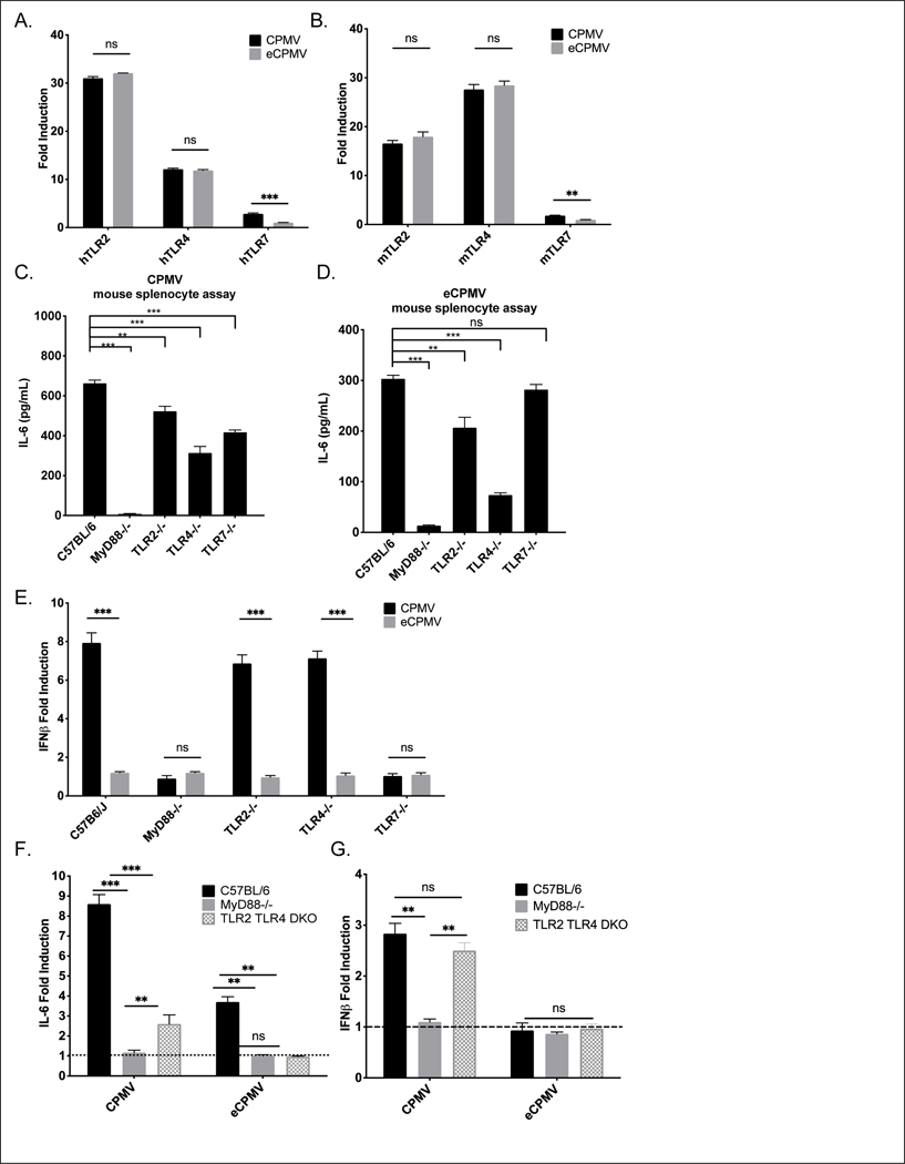
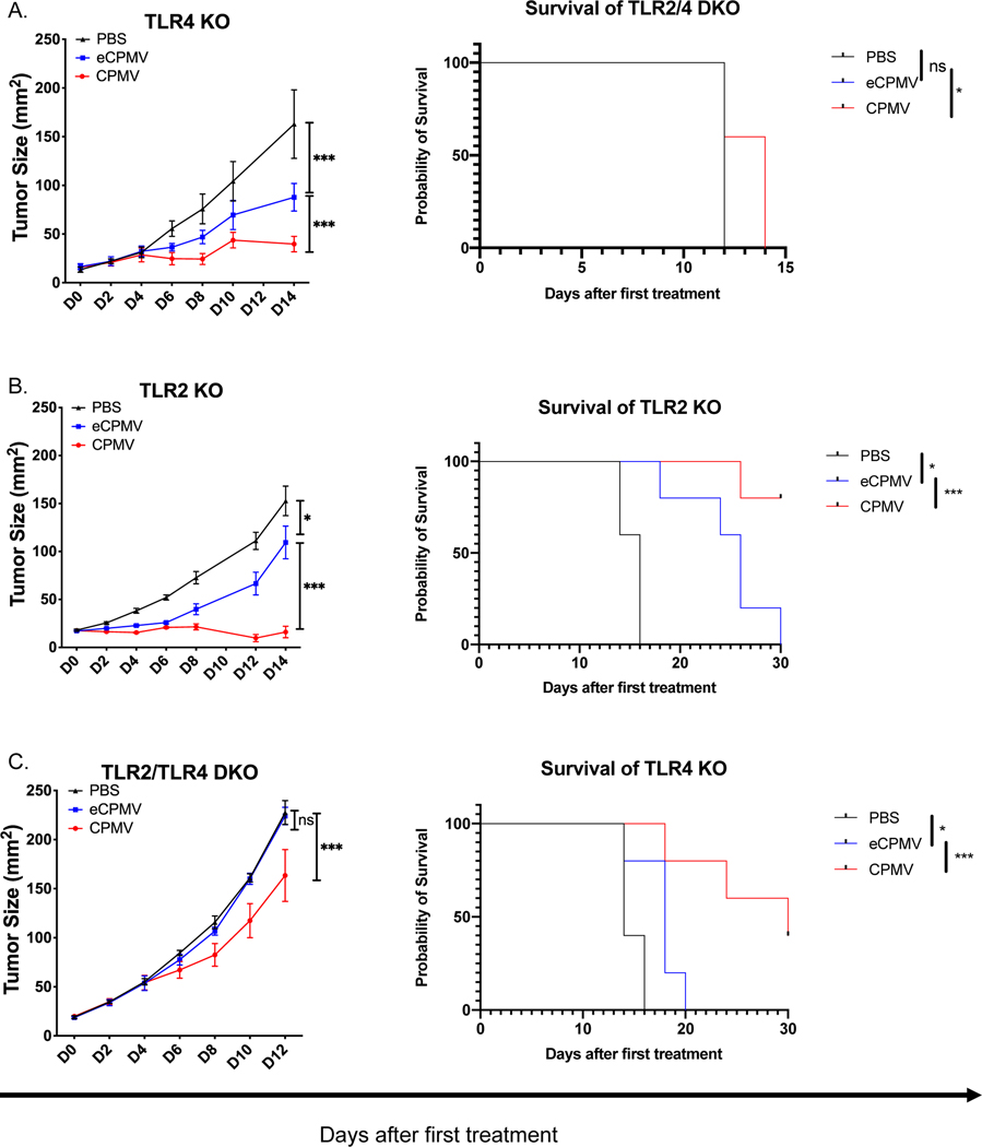
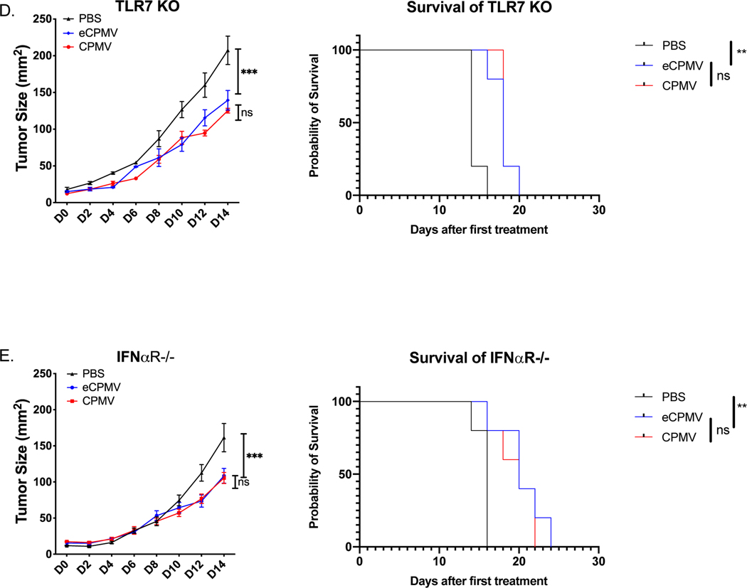
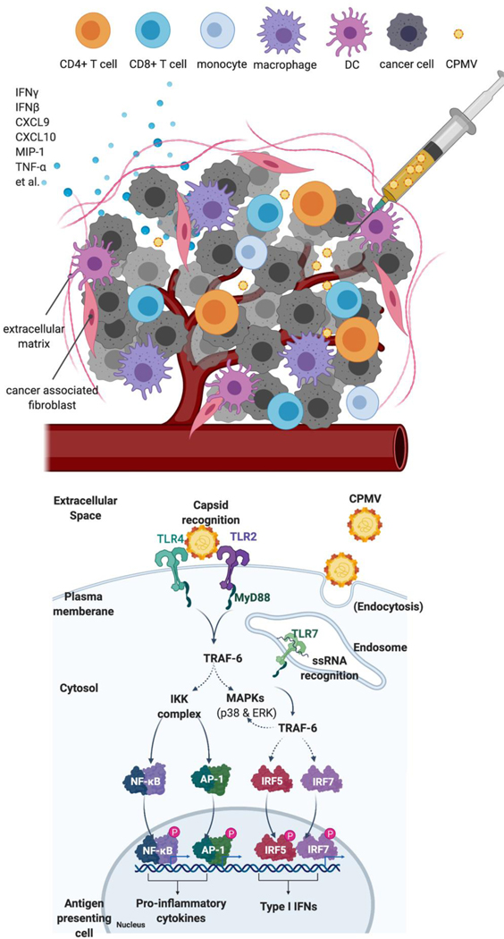
Cowpea mosaic virus (CPMV), a non-enveloped plant virus, and empty CPMV (eCPMV), a virus-like particle (VLP) composed of CPMV capsid without nucleic acids, are potent in situ cancer vaccines when administered intratumorally (I.T.). However, it is unclear how immune cells recognize these nanoparticles and why they are immunogenic, which was investigated in this study. CPMV generated stronger selective induction of cytokines and chemokines in naïve mouse splenocytes and exhibited more potent anti-tumor efficacy than eCPMV. MyD88 is required for both CPMV- and eCPMV-elicited immune responses. Screening with human embryonic kidney (HEK)-293 cell toll-like receptor (TLR) reporter assays along with experiments in corresponding TLR-/- mice indicated CPMV and eCPMV capsids are recognized by MyD88-dependent TLR2 and TLR4. CPMV, but not eCPMV, is additionally recognized by TLR7. Secretion of type I interferons (IFNs), which requires the interaction between TLR7 and encapsulated single-stranded RNAs (ssRNAs), is critical to CPMV's better efficacy. The same recognition mechanisms are also functional in human peripheral blood mononuclear cells (PBMCs). Overall, these findings link CPMV immunotherapy efficacy with molecular recognition, provide rationale for how to develop more potent viral particles, accentuate the value of multi-TLR agonists as in situ cancer vaccines, and highlight the functional importance of type I IFNs for in situ vaccination.

Keywords: Cancer immunotherapy; Cowpea mosaic virus; In situ vaccination; Nanoparticle; Toll-like receptors; Virus-like nanoparticle.


Conflict of interest statement

Declaration of competing Interest:

Drs. Fiering and Steinmetz are co-founders of and have a financial relationship with Mosaic Immunoengineering Inc, which may be considered potential competing interest. The other authors declare no potential conflict of interest.

Figures

Fig 1.
Fig 1.. MyD88 is required for CPMV and eCPMV-elicited upregulation of cytokines and chemokines in vitro and anti-tumor immunity in vivo.
A. Schematic of splenocyte assay design. Splenocytes were cultured for 24hr with (e)CPMV and cytokine levels were analyzed. B. Induction of cytokines and chemokines by CPMV and eCPMV did not occur in MyD88−/− mouse splenocytes and IFNβ was only induced by CPMV (n=3). C. Schematic of in situ vaccination strategy. 1.25×10⁵ of B16F10 melanoma cells were injected intradermally on day −7. 100 ug of CPMV or eCPMV was injected intratumorally on D0 and D7. Tumor growth was monitored every other day till 30 days after first treatment and euthanized or when the tumor size reached 200mm2. D-F. MyD88, but not TRIF, is required for CPMV and eCPMV-elicited anti-tumor immunity in vivo (n=5). Data for bar graphs calculated using unpaired Student’s t-test. Tumor progression was monitored by measuring tumor surface area (length x width). Growth curves were analyzed using two-way ANOVA. survival curves were analyzed using log-rank (Mantel-Cox) test, with p>0.05 as ns, p <0.05 as *, p <0.01 as **, and p <0.001 as ***. All experiments are repeated at least once with similar results.
Fig 2.
Fig 2.. In vitro, both CPMV and eCPMV are recognized by TLR2 and TLR4; however, CPMV is recognized only by TLR 7 to induce type I IFNs.
A, B. Screened by hTLR and mTLR HEK-Blue reporter cells (n=3), both CPMV and eCPMV showed induction of TLR2 and TLR4 transactivation; however, only CPMV shows induction of TLR7 transactivation. C, D. Knockout of either TLR2, TLR4 or TLR7 in mouse splenocytes (n=3) was reduced but did not completely eliminate induction of IL-6. E. Type I IFNs are only induced by CPMV but not eCPMV in a TLR7-dependent manner in mouse splenocyte assays (n=3). F, G. Knockout of both TLR2 and TLR4 in mouse splenocytes eliminated IL-6 induced by eCPMV and attenuated IL-6 induction but not IFNβ induction by CPMV (n=3). Data for bar graphs calculated using unpaired Student’s t-test with p>0.05 as ns, p <0.05 as *, p <0.01 as **, and p <0.001 as ***. All experiments are repeated at least once with similar results.
Fig 3.
Fig 3.. TLR2 and TLR4 are complementary to each other for the antitumor efficacy by CPMV and eCPMV in vivo; ssRNAs in CPMV induce type I IFNs by TLR7 which improves efficacy.
ISV strategy to treat B16F10 intradermal tumors as shown in Fig. 1c. A, B. CPMV and eCPMV remained normally effective in TLR2 KO or TLR4 KO mice (n=5). C. Knockout of both TLR2 and TLR4 attenuated antitumor efficacy of CPMV and eliminated the antitumor efficacy of eCPMV. D, E. CPMV and eCPMV have equal antitumor efficacy in TLR7 KO and in IFNαR KO mice (n=5). Tumor progression was monitored by measuring tumor area (length x width). Tumor progression was monitored by measuring tumor surface area (length x width). Growth curves were analyzed using two-way ANOVA. survival curves were analyzed using log-rank (Mantel-Cox) test, p <0.05 as *, p <0.01 as **, and p <0.001 as ***. All experiments are repeated at least once with similar results.
Fig 4.
Fig 4.. Human PBMC recognized and responded to CPMV and eCPMV in a similar manner as mouse splenocytes.
A. Selective upregulation of cytokines and chemokines by CPMV and eCPMV in human PBMCs compared to control, and IFNα was only induced by CPMV (n=3). B, C. Only blocking both TLR2 and TLR4 by antibodies could eliminate IL-6 induced by eCPMV/ attenuated IL-6 induced but not IFNβ by CPMV in human PBMC assay (data is the average of results from n=3 different PBMC donors). Data for bar graphs calculated using unpaired Student’s t-test with p>0.05 as ns, p <0.05 as *, p <0.01 as **, and p <0.001 as ***. All experiments are repeated at least once with similar results.
Fig. 5.
Fig. 5.. Improved efficacy is observed in the combination of eCPMV and R848.
A. Schematic of in situ vaccination strategy adapted from Fig 1c. 1.25×10⁵ of B16F10 melanoma cells were injected intradermally on day −7. 100 ug of CPMV, eCPMV, or eCPMV + 40 ug of R848 was injected intratumorally on D0 and D7. Tumor growth was monitored every other day. B, C. ISV treatment with combination of eCPMV (100 ug) and R848 (40 ug) showed improved efficacy but is still inferior to CPMV (100 ug) in B16F10-tumor bearing mice (n=5). Tumor progression was monitored by measuring tumor surface area (length x width). Growth curves were analyzed using two-way ANOVA. survival curves were analyzed using log-rank (Mantel-Cox) test, p <0.05 as *, p <0.01 as **, and p <0.001 as ***. All experiments are repeated at least once with similar results.
Fig 6:
Fig 6:. Schematic illustration of CPMV recognition by TLR2, TLR4 and TLR7.
Upper panel: CPMV is i.t. injected into a solid tumor, which is recognized by immune cells and stimulates proinflammatory cytokines and chemokines to recruit other immune cells and stimulate antitumor immunity. Lower panel: One APC (like dendritic cell) in the injected site is shown: the capsid of CPMV and eCPMV (not shown) is recognized by TLR2 and TLR4 located at plasma membrane. Upon entry by endocytosis, ssRNA of CPMV is recognized by TLR7. Downstream signaling cascade following TLR2/TLR4, and TLR7 activation is MyD88, which is the adaptor protein that initiates the different pathways activating IRF5, IRF7 (plasmacytoid DCs only), NF-κB, and AP-1 in order to induce the antiviral response. NF-κB and AP-1 combine to induce the transcription of inflammatory cytokines. IRF5/IRF7 in association with both NF-κB and AP-1 form the enhanceosome for the transcription of IFN-β/IFN-α, respectively. In addition, IRF7 also has IFN-β-stimulatory effect.

References

    1. Rabinovich GA, Gabrilovich D, & Sotomayor EM (2007). Immunosuppressive strategies that are mediated by tumor cells. Annual review of immunology, 25, 267–296. 10.1146/annurev.immunol.25.022106.141609 - DOI - PMC - PubMed
    1. Dougan M, & Dranoff G. (2009). Immune therapy for cancer. Annual review of immunology, 27, 83–117. 10.1146/annurev.immunol.021908.132544 - DOI - PubMed
    1. Tang J, Yu JX, Hubbard-Lucey VM, Neftelinov ST, Hodge JP, & Lin Y. (2018). Trial watch: The clinical trial landscape for PD1/PDL1 immune checkpoint inhibitors. Nature reviews. Drug discovery, 17(12), 854–855. 10.1038/nrd.2018.210 - DOI - PubMed
    1. Lu J, Lee-Gabel L, Nadeau MC, Ferencz TM, & Soefje SA (2015). Clinical evaluation of compounds targeting PD-1/PD-L1 pathway for cancer immunotherapy. Journal of oncology pharmacy practice : official publication of the International Society of Oncology Pharmacy Practitioners, 21(6), 451–467. 10.1177/1078155214538087 - DOI - PubMed
    1. Yun S, Vincelette ND, Green MR, Wahner Hendrickson AE, & Abraham I. (2016). Targeting immune checkpoints in unresectable metastatic cutaneous melanoma: a systematic review and meta-analysis of anti-CTLA-4 and anti-PD-1 agents trials. Cancer medicine, 5(7), 1481–1491. 10.1002/cam4.732 - DOI - PMC - PubMed

Publication types

Substances