. 2021 Jun 14;4(8):e202001000.
doi: 10.26508/lsa.202001000. Print 2021 Aug.

IL-13 deficiency exacerbates lung damage and impairs epithelial-derived type 2 molecules during nematode infection

Affiliations

IL-13 deficiency exacerbates lung damage and impairs epithelial-derived type 2 molecules during nematode infection

Alistair L Chenery et al. Life Sci Alliance. .

Abstract

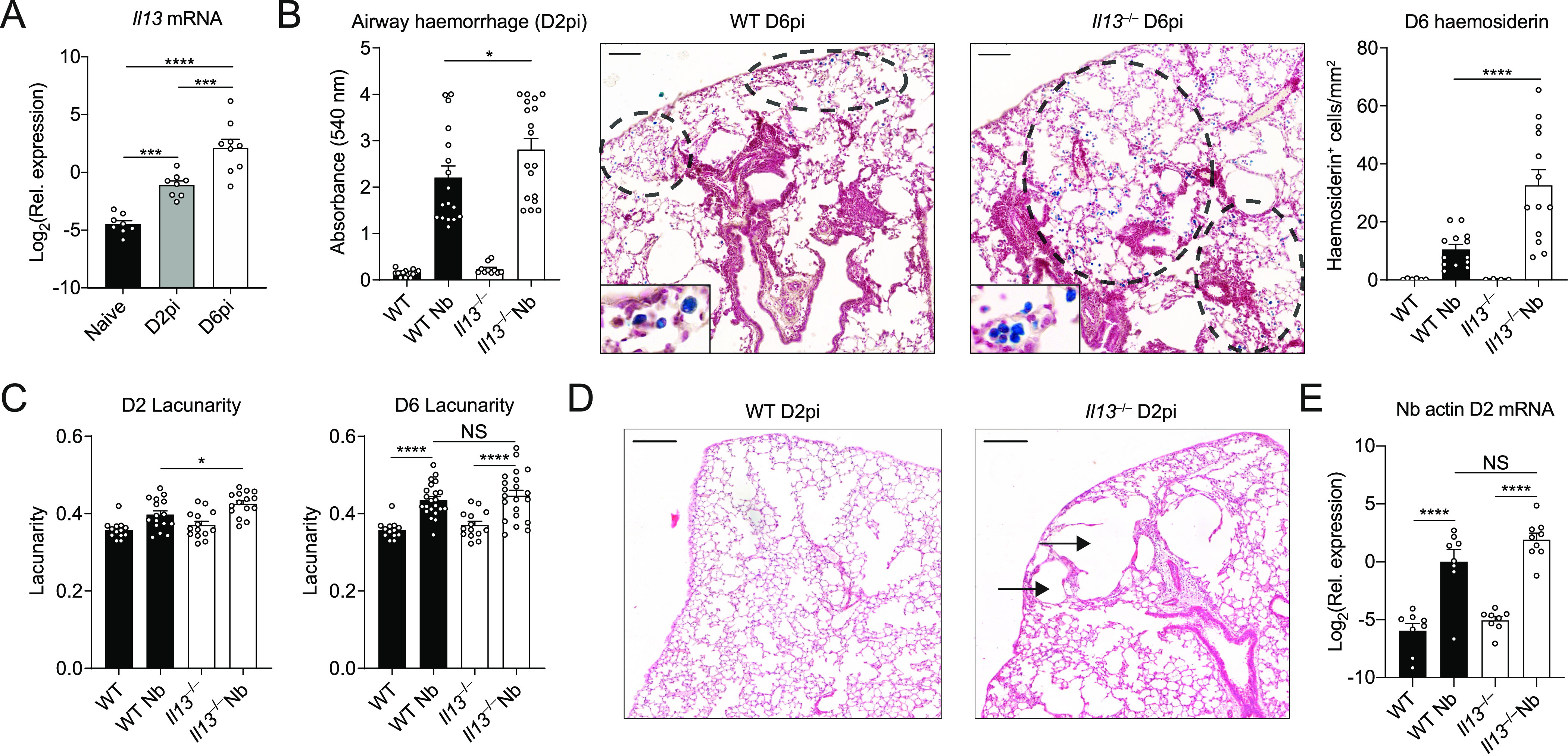
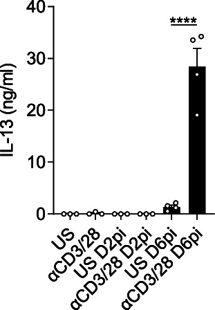
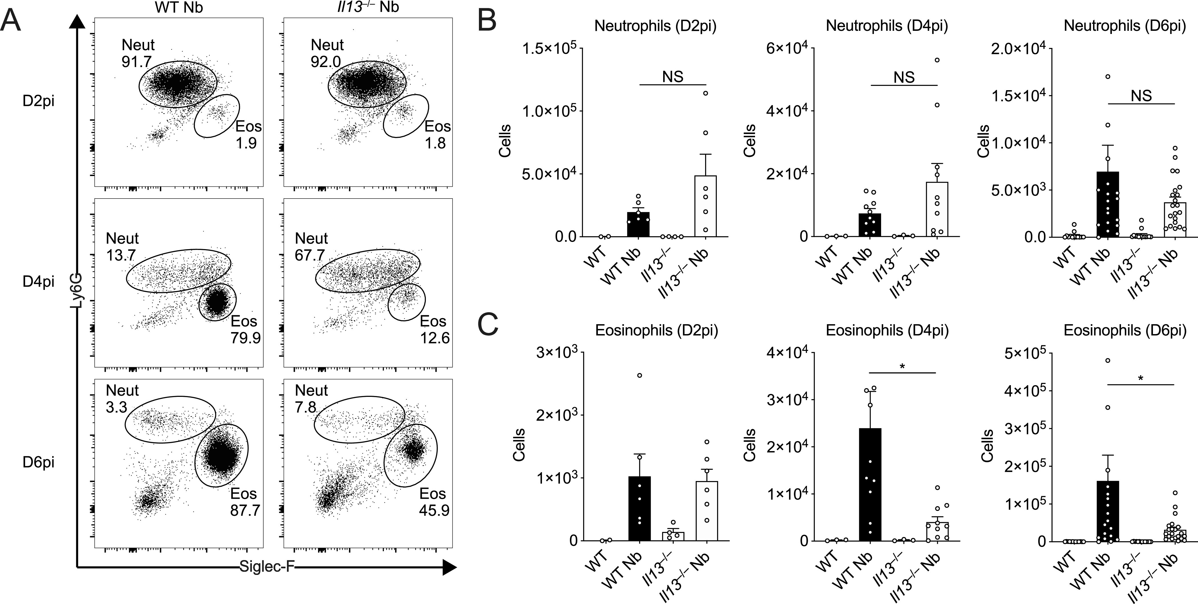
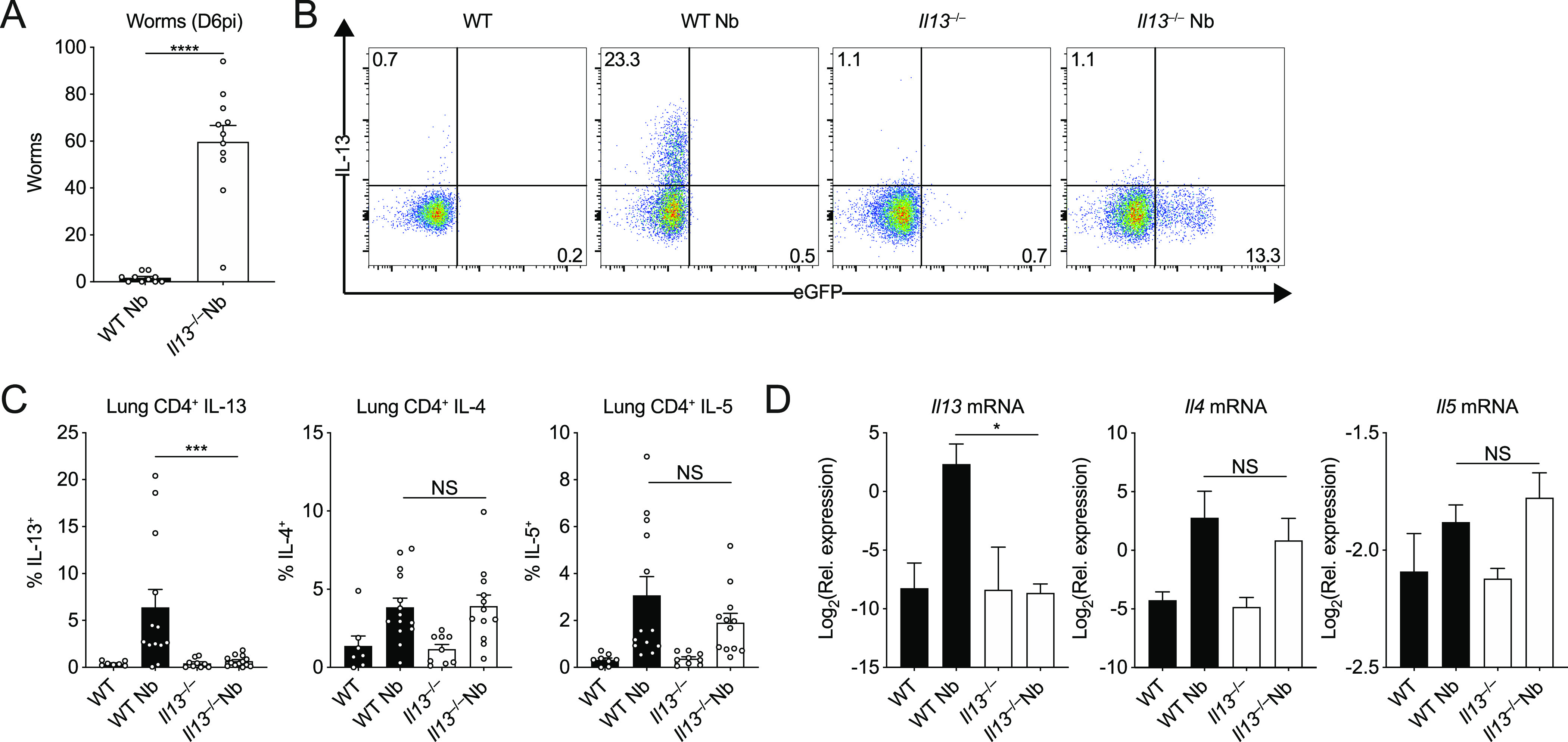
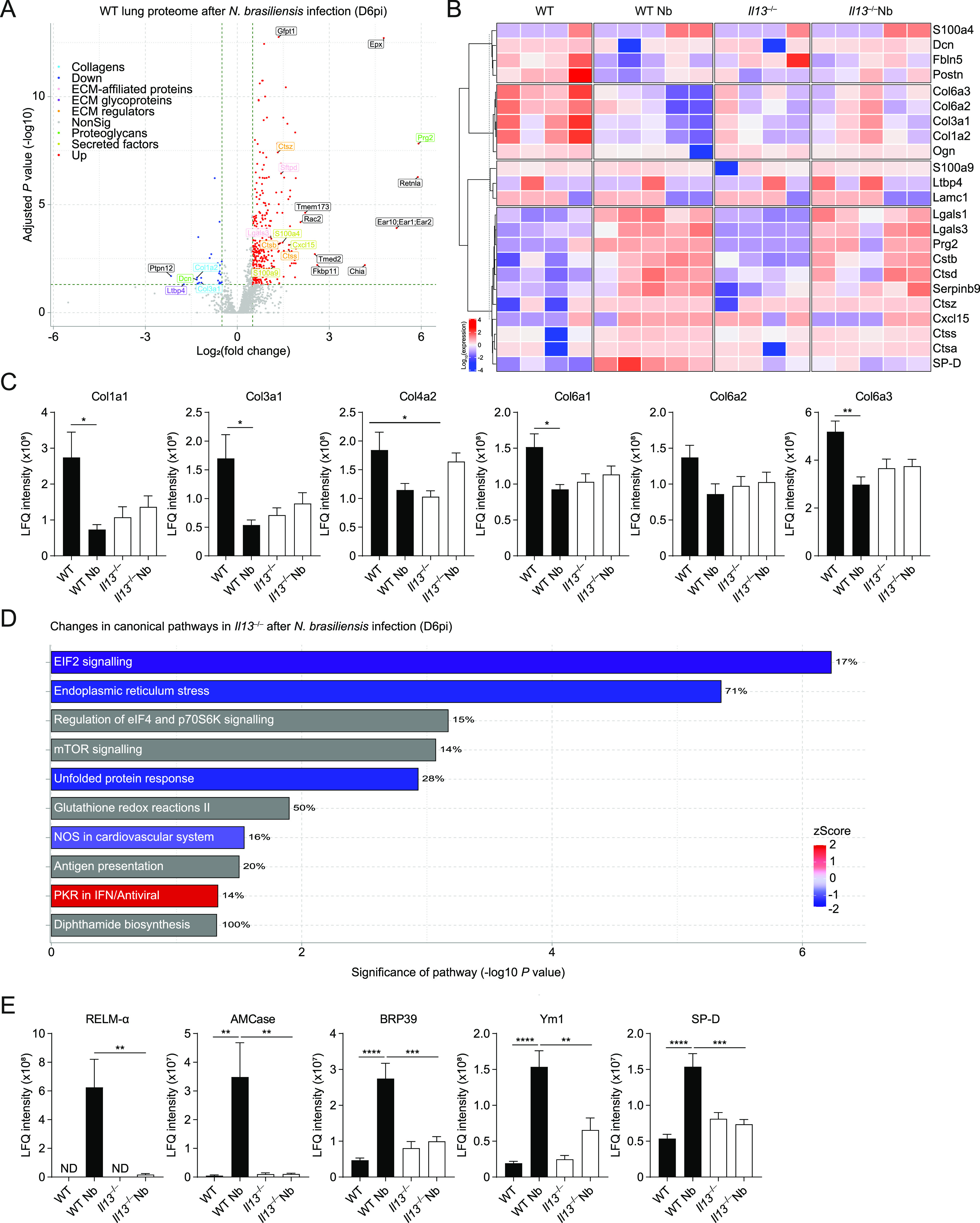
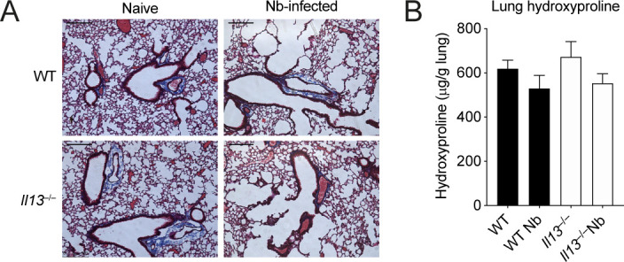
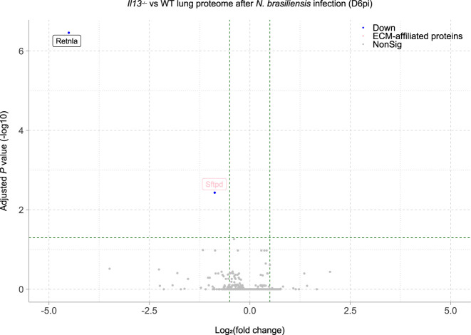
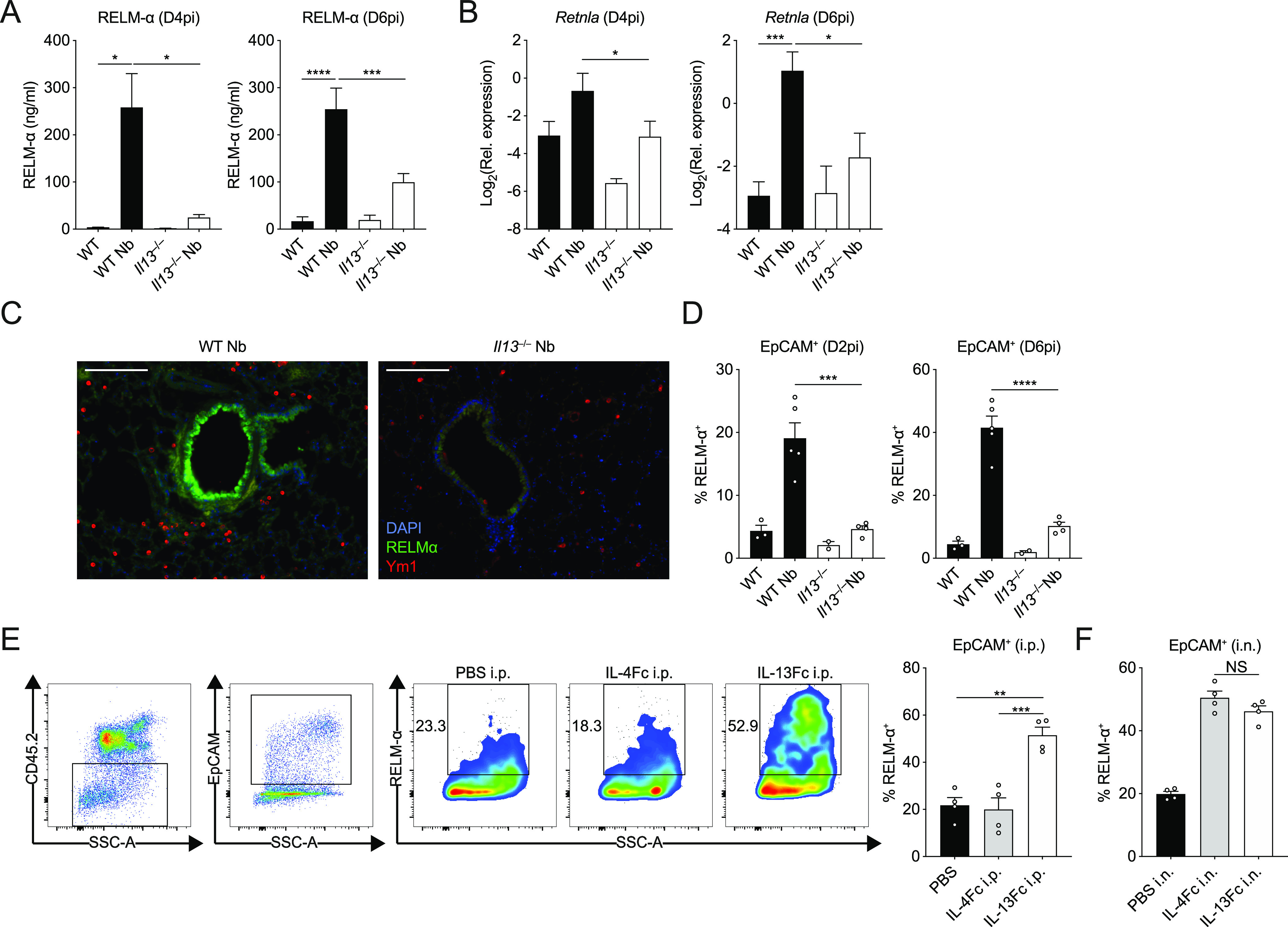
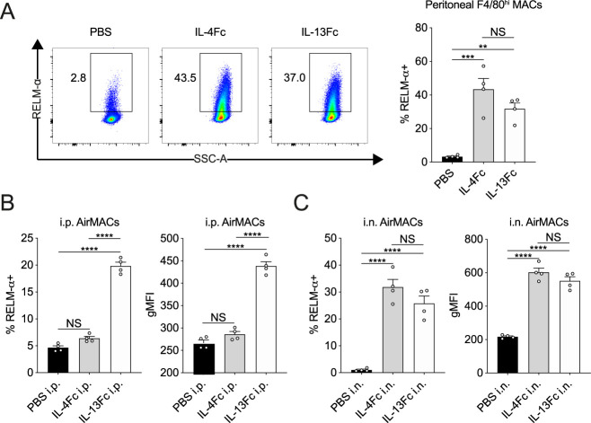
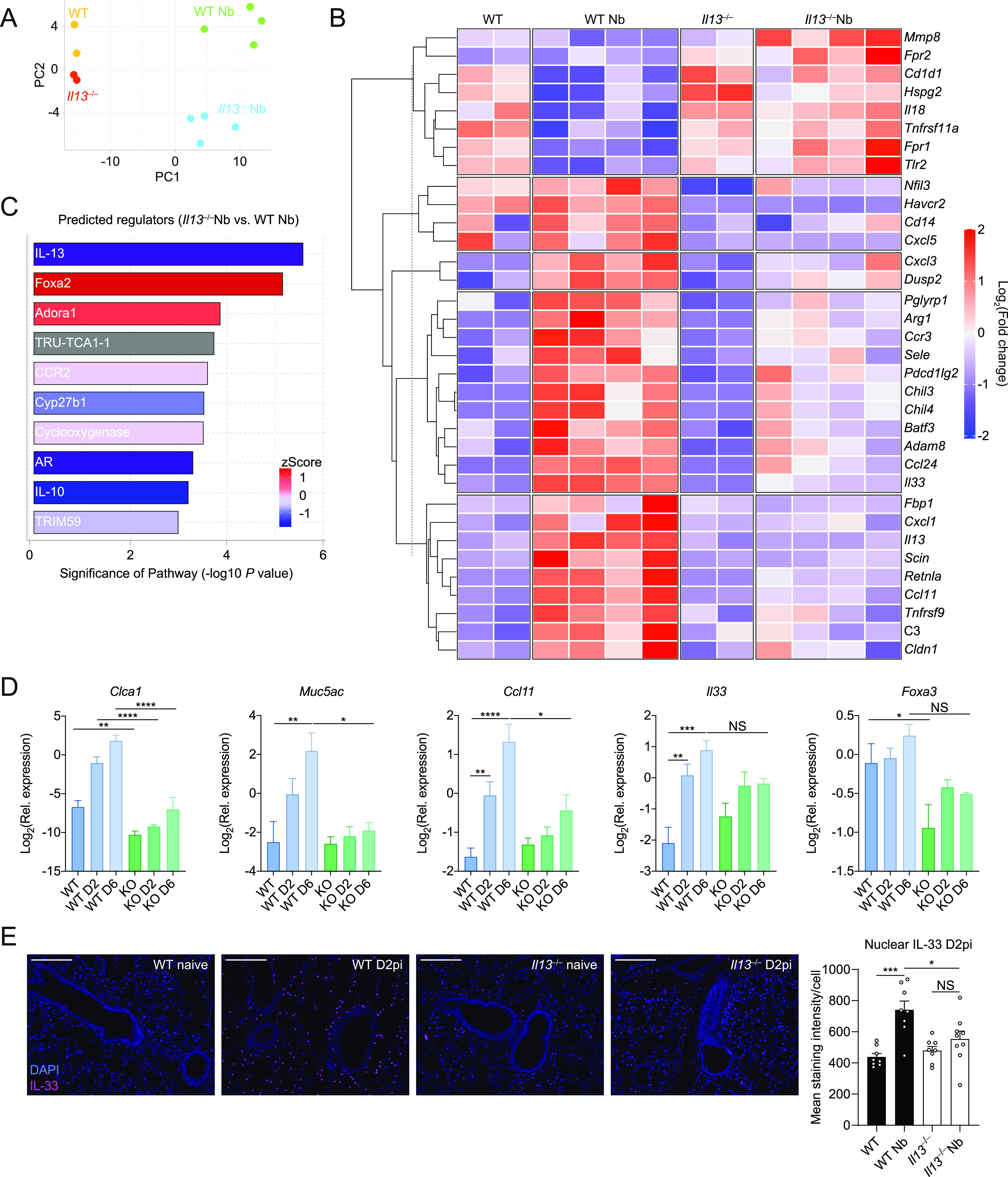
IL-13 is implicated in effective repair after acute lung injury and the pathogenesis of chronic diseases such as allergic asthma. Both these processes involve matrix remodelling, but understanding the specific contribution of IL-13 has been challenging because IL-13 shares receptors and signalling pathways with IL-4. Here, we used Nippostrongylus brasiliensis infection as a model of acute lung damage comparing responses between WT and IL-13-deficient mice, in which IL-4 signalling is intact. We found that IL-13 played a critical role in limiting tissue injury and haemorrhaging in the lung, and through proteomic and transcriptomic profiling, identified IL-13-dependent changes in matrix and associated regulators. We further showed a requirement for IL-13 in the induction of epithelial-derived type 2 effector molecules such as RELM-α and surfactant protein D. Pathway analyses predicted that IL-13 induced cellular stress responses and regulated lung epithelial cell differentiation by suppression of Foxa2 pathways. Thus, in the context of acute lung damage, IL-13 has tissue-protective functions and regulates epithelial cell responses during type 2 immunity.


Conflict of interest statement

The authors declare that they have no conflict of interest.

Figures

Figure 1.
Figure 1.. Tissue-protective role for IL-13 during acute lung injury.
WT and Il13−/− mice were infected with Nippostrongylus brasiliensis (Nb). (A) On day 2 post-infection (D2pi) and D6pi, Il13 mRNA levels were measured in the lungs of WT mice by quantitative real-time PCR (data normalised against housekeeping gene Rpl13a). (B) On D2pi, BAL fluid absorbance at 540 nm was quantified to measure airway haemorrhage. On D6pi, lung lobe sections were stained with Prussian blue and haemosiderin-laden cells (blue) were enumerated per area of tissue (scale bar = 100 µm). (C) To measure airway damage on D2pi and D6pi, lacunarity for whole lung lobes was computed. (D) Representative haematoxylin and eosin images of infected WT and Il13−/− lungs showing alveolar damage in the tissue (scale bar = 200 µm). (E) Nb-specific actin mRNA in lung tissue was measured on D2pi by quantitative real-time PCR (data normalised against housekeeping gene Rpl13a). Data (mean ± SEM) were pooled from three individual experiments with three to six mice per group (per experiment). NS not significant, *P < 0.05, ****P < 0.0001 (one-way ANOVA and Tukey–Kramer post hoc test).
Figure S1.
Figure S1.. Measurement of IL-13 after Nippostrongylus brasiliensis infection.
Cell homogenates of lungs from either naïve, day 2 post-infection, and D6pi WT mice were either unstimulated (US) or stimulated with anti-CD3/CD28 for 72 h and IL-13 levels were measured in the supernatants by ELISA. Data (mean ± SEM) are from a single experiment at different time points with three to four mice per group.
Figure 2.
Figure 2.. IL-13-dependent airway eosinophilia during Nippostrongylus brasiliensis infection.
WT and Il13−/− mice were infected with N. brasiliensis (Nb) and BAL cells were analysed by flow cytometry. (A) Representative plots of percentages of BAL CD11cCD11b+Ly6G+ neutrophils and CD11cCD11b+Siglec-F+ eosinophils on D2, D4, and D6pi. Numbers indicate percentage of cells within total live CD45.2+ cells. (B, C) Total BAL neutrophil and (C) eosinophil cell counts on D2, D4, and D6pi. Data (mean ± SEM) were representative (day 2 post-infection) or pooled (D4 and D6pi) from four individual experiments with three to five mice per group (per experiment). NS: not significant, *P < 0.05 (one-way ANOVA and Tukey–Kramer post hoc test).
Figure 3.
Figure 3.. IL-4 and IL-5 do not compensate in the absence of IL-13 during Nippostrongylus brasiliensis infection.
WT and Il13−/− mice were infected with N. brasiliensis (Nb). (A) On D6pi, adult worms in the small intestine were quantified. (B) Representative flow cytometry plots of lung CD4+ T cells stimulated ex vivo to measure intracellular WT IL-13 and KO eGFP expression. (C) Percentages of CD4+ T cells expressing IL-13, IL-4, and IL-5. (D) Whole lung Il13, Il4, and Il5 mRNA was measured by quantitative real-time PCR (data normalised against housekeeping gene Rpl13a). Data (mean ± SEM) were pooled from three individual experiments with three to five mice per group (per experiment). NS: not significant, *P < 0.05, ***P < 0.001, ****P < 0.0001 (one-way ANOVA and Tukey–Kramer post hoc test).
Figure 4.
Figure 4.. IL-13–dependent lung proteomic changes during Nippostrongylus brasiliensis infection.
WT and Il13−/− mice were infected with N. brasiliensis (Nb) and on D6pi lungs were prepared for proteomic analysis. (A) Volcano plot of infected WT lungs (D6pi) showing differential expression of up- (red) or down- (blue) regulated proteins with matrisome annotations (fold change relative to naïve WT mice). Black labels indicate proteins lacking matrisome annotations. (B) Unsupervised, hierarchically clustered heatmap of expression of matrisome proteins comparing naïve and Nb-infected groups of WT and Il13−/− mice on D6pi. (C) Columns in each set represent different (biological repeat) mice in each group (C) Relative abundance (label-free quantification [LFQ] intensity) of collagen peptides comparing infected WT and Il13−/− lungs on D6pi. (D) Predicted changes in canonical pathways based on changes in the proteome of infected Il13−/− mice compared with infected WT mice (down-regulation in blue, up-regulation in red, and no specified direction in grey). Percentage indicates the relative number of proteins that are regulated in each pathway. (E) Relative abundance of peptides highly associated with type 2 immunity comparing infected WT and Il13−/− lungs on D6pi. Data (mean ± SEM in C and E) are pooled from two independent mass spectrometry runs with four to five mice per group in total. *P < 0.05, **P < 0.01, ***P < 0.001, ****P < 0.0001 (one-way ANOVA and Tukey–Kramer post hoc test).
Figure S2.
Figure S2.. Analysis of total collagen in the lungs of Il13−/− mice after Nippostrongylus brasiliensis infection on D6pi.
(A) Lung sections were prepared and stained with Masson’s trichrome stain and imaged to visualise collagen (scale bar = 200 µm). (B) Hydroxyproline levels from whole lungs were measured. (A, B) Data are representative (A) or pooled (mean ± SEM in B) from two individual experiments with four to five mice per group (per experiment).
Figure S3.
Figure S3.. Volcano plot of differentially expressed proteins (by adjusted P-value) in the lungs of Nippostrongylus brasiliensis–infected Il13−/− mice compared with infected WT mice on D6pi.
Figure 5.
Figure 5.. Lung epithelial cell expression and airway release of RELM-α is IL-13 dependent.
WT and Il13−/− mice were infected with Nippostrongylus brasiliensis (Nb). (A) On D4 and D6pi RELM-α protein levels in the BAL fluid were measured by ELISA. (B) D4 and D6pi whole lung Retnla mRNA was measured by quantitative real-time PCR (data normalised against housekeeping gene Rpl13a). (C) Lung RELM-α (green) and Ym1 (red) were imaged by immunofluorescence microscopy (scale bar = 100 µm). (D) On D2 and D6pi, CD45EpCAM+ lung epithelial cells were analysed and quantified by flow cytometry to measure intracellular RELM-α. (E, F) WT mice were injected with either PBS, IL-4Fc, or IL-13Fc i.p. or (F) i.n. and 18 h later, CD45EpCAM+ lung epithelial cell RELM-α expression was measured by flow cytometry. (A, B) Data (mean ± SEM) in (A, B) were pooled from three individual experiments with three to five mice per group (per experiment). (C, D, E, F) Data (mean ± SEM) in (C, D, E, F) were representative of two individual experiments with two to five mice per group (per experiment). NS: not significant, *P < 0.05, **P < 0.01, ***P < 0.001, ****P < 0.0001 (one-way ANOVA and Tukey–Kramer post hoc test).
Figure S4.
Figure S4.. RELM-α expression in macrophages after IL-13 or IL-4 delivery.
(A) Peritoneal resident (F4/80hi) macrophage expression of RELM-α after intraperitoneal injection of either PBS, IL-4Fc, or IL-13Fc after 18 h. (B, C) Comparison of airway macrophage (CD11c+Siglec-F+) expression of RELM-α after either (B) intraperitoneal or (C) intranasal delivery of either PBS, IL-4Fc, or IL-13Fc. Data (mean ± SEM) were representative of two individual experiments with two to five mice per group (per experiment). NS, not significant, ****P < 0.0001 (one-way ANOVA and Tukey–Kramer post hoc test).
Figure 6.
Figure 6.. Transcriptional profiling of IL-13–dependent genes in the lung after Nippostrongylus brasiliensis infection.
Whole lung RNA from WT and Il13−/− mice infected with N. brasiliensis (Nb) on D6pi was analysed by Nanostring. (A) Principle components analysis of naïve and infected WT and Il13−/− mice. (B) Unsupervised, hierarchically clustered heat map of genes differentially expressed between mouse groups with fold change expression level indicated by colour. (C) Columns in each set represent different (biological repeat) mice in each group (C) Predicted upstream regulators from Ingenuity Pathway Analysis. (D) Expression of Foxa2-regulated genes Clca1, Muc5ac, Ccl11, Il33, and Foxa3 were measured in lung tissues on day 2 post-infection and D6pi by quantitative real-time PCR (data normalised against housekeeping gene Rpl13a). (E) Immunofluorescence staining of nuclear IL-33 (magenta) in the parenchyma of the lung and quantification of mean integrated density (IntDen) (scale bar = 100 µm). Data in (A, B, C) are from a single Nanostring run with samples from two to four mice per group. Data (mean ± SEM) in (D, E) were pooled from two individual experiments with three to five mice per group (per experiment). NS, not significant, *P < 0.05, **P < 0.01, ***P < 0.001, ****P < 0.0001 (one-way ANOVA and Tukey–Kramer post hoc test).

References

    1. Bagnasco D, Ferrando M, Varricchi G, Passalacqua G, Canonica GW (2016) A critical evaluation of Anti-IL-13 and Anti-IL-4 strategies in severe asthma. Int Arch Allergy Immunol 170: 122–131. 10.1159/000447692 - DOI - PubMed
    1. Bao K, Reinhardt RL (2015) The differential expression of IL-4 and IL-13 and its impact on type-2 immunity. Cytokine 75: 25–37. 10.1016/j.cyto.2015.05.008 - DOI - PMC - PubMed
    1. Barner M, Mohrs M, Brombacher F, Kopf M (1998) Differences between IL-4R alpha-deficient and IL-4-deficient mice reveal a role for IL-13 in the regulation of Th2 responses. Curr Biol 8: 669–672. 10.1016/s0960-9822(98)70256-8 - DOI - PubMed
    1. Bouchery T, Kyle R, Camberis M, Shepherd A, Filbey K, Smith A, Harvie M, Painter G, Johnston K, Ferguson P, et al. (2015) ILC2s and T cells cooperate to ensure maintenance of M2 macrophages for lung immunity against hookworms. Nat Commun 6: 6970. 10.1038/ncomms7970 - DOI - PubMed
    1. Castro M, Corren J, Pavord ID, Maspero J, Wenzel S, Rabe KF, Busse WW, Ford L, Sher L, FitzGerald JM, et al. (2018) Dupilumab efficacy and safety in moderate-to-severe uncontrolled asthma. N Engl J Med 378: 2486–2496. 10.1056/NEJMoa1804092 - DOI - PubMed

Publication types

Substances

LinkOut - more resources