. 2021 Jun 14;12(1):3612.
doi: 10.1038/s41467-021-23848-9.

SARS-CoV-2 infection and transmission in the North American deer mouse

Affiliations

SARS-CoV-2 infection and transmission in the North American deer mouse

Bryan D Griffin et al. Nat Commun. .

Abstract

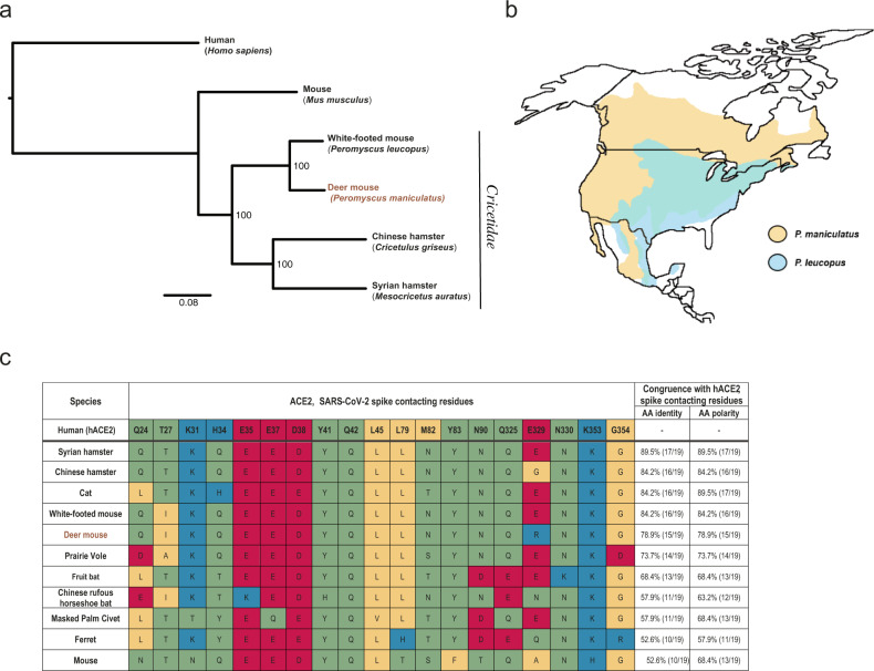
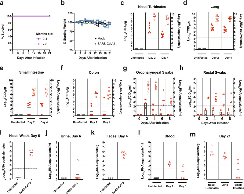
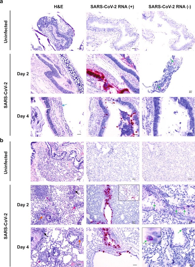
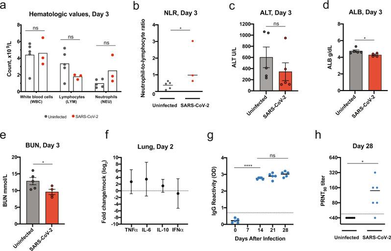
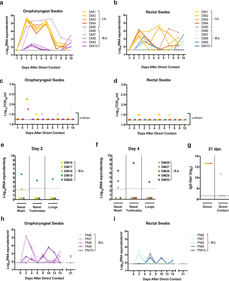
Widespread circulation of SARS-CoV-2 in humans raises the theoretical risk of reverse zoonosis events with wildlife, reintroductions of SARS-CoV-2 into permissive nondomesticated animals. Here we report that North American deer mice (Peromyscus maniculatus) are susceptible to SARS-CoV-2 infection following intranasal exposure to a human isolate, resulting in viral replication in the upper and lower respiratory tract with little or no signs of disease. Further, shed infectious virus is detectable in nasal washes, oropharyngeal and rectal swabs, and viral RNA is detectable in feces and occasionally urine. We further show that deer mice are capable of transmitting SARS-CoV-2 to naïve deer mice through direct contact. The extent to which these observations may translate to wild deer mouse populations remains unclear, and the risk of reverse zoonosis and/or the potential for the establishment of Peromyscus rodents as a North American reservoir for SARS-CoV-2 remains unknown.


Conflict of interest statement

The authors declare no conflicts of interest.

Figures

Fig. 1
Fig. 1. Deer mouse range and predicted susceptibility to SARS-CoV-2.
a Phylogeny showing the relationships among selected members of the Cricetidae family with mice and humans. b Geographical distribution of deer mice (based on data from Hall, 1981). c Alignment of human ACE2 (hACE2) amino acid residues known to confer efficient binding of the RBD of SARS-CoV-2 spike with the corresponding ACE2 amino acid residues from selected members of the Cricetidae family and other naturally or experimentally susceptible host species. The physiochemical properties of the amino acids residues are indicated: nonpolar (yellow), polar (green), acidic (red), and basic residues (blue).
Fig. 2
Fig. 2. SARS-CoV-2 infection of adult deer mice.
a–m Eight to thirty two-week old female or male deer mice (P. maniculatus) were inoculated with 105 TCID50 or 106 TCID50 of SARS-CoV-2 by an intranasal route (i.n.) of administration and compared with age-matched uninfected controls. A summary table outlining the experimental design for the infection studies with reference to the corresponding figure panels is provided (Supplementary Table. 1). Solid lines indicate mean, error bars indicate SD. Dashed lines and dotted lines indicate the limit of detection for the TCID50 assay and qRT-PCR assay, respectively. a, b, Kaplan–Meier curve depicting survival data (a) and weight data (b) over the course of 21 days following SARS-CoV-2 exposure (challenge dose, 105 TCID50). cf, Infectious viral load (filled in circles, left axis) and vRNA levels (empty circles, right axis) in the (c) nasal turbinates, (d) lung, (e) small intestine, and (f) colon (challenge dose, 105 TCID50). g, h, Infectious viral load (filled in circles with bars, left axis) and viral RNA levels (empty circles, right axis) in (g) oropharyngeal swabs and (h) rectal swab samples (challenge dose, 105 TCID50). i, Viral RNA levels in nasal washes at 6 dpi (infectious dose, 105 TCID50). The sample was obtained for 4/5 deer mice. jl, Viral RNA levels in the (j) urine, (k) feces, and (l) blood at the indicated times postinfection (infectious dose, 106 TCID50). m, Viral RNA levels in the nasal turbinates, lung, and small intestines at 21 days postinfection (infectious dose, 105 TCID50). Data were collected from two independent experiments. (a, b, n = 6; cf, n = 3/6, for uninfected/infected; g, h, n = 5/6; ik, n = 3/4 (i) or 3/5 (j, k), for uninfected/infected; l, n = 5 (uninfected, 1 dpi) or 4 (3 dpi); m, n = 4 biologically independent samples (individual animals) were examined).
Fig. 3
Fig. 3. Histopathology and virus distribution.
Hematoxylin/eosin (H&E) staining (left) and in situ hybridization (ISH) using antisense probes that detect the SARS-CoV-2 genome/mRNA (middle) and sense probes that detect anti-genomic RNA (right) were carried out on (a) nasal turbinates and (b) lung tissue of uninfected and SARS-CoV-2-infected deer mice (105 TCID50 i.n. route) at 2 and 4 dpi. Positive detection of viral genomic RNA/mRNA or anti-genomic RNA is indicated by magenta staining (middle/right panels and insets). Arrows indicate perivascular infiltrations of histiocytes and neutrophils (black), peribronchiolar infiltrations of histiocytes and neutrophils (orange), mild neutrophilic infiltration in the submucosa (blue), occasionally observed discrete foci of interstitial pneumonia (purple), occasionally observed multinucleated syncytial cells (magenta), and anti-genomic RNA occasionally in individual scattered bronchiolar epithelial cells (green and right inset). The magnification is ×20 for H&E and ×20 for ISH unless otherwise indicated. Scale bars =100 μm, 50 μm, and 20 μm for 10×, 20×, and 40×, respectively. A total of three biological replicates (individual animals) were assessed at each time point, and the selected panels are representative of these findings.
Fig. 4
Fig. 4. Deer mouse host response to SARS-CoV-2 infection.
a–e Hematological levels and serum biochemistry were measured in uninfected and SARS-CoV-2-infected deer mice (infectious dose 106 TCID50, i.n. route) at 3 dpi, including (a) white blood cell, lymphocyte, and neutrophil counts, (b) the neutrophil-to-lymphocyte ratio, (c) alanine aminotransferase (ALT), (d) blood albumin (ALB), and (e) blood urea nitrogen (BUN). f Cytokine gene expression was measured for TNFα, IL-6, IL-10, and IFNα in the lungs of SARS-CoV-2-infected deer mice and displayed relative to age-matched mock-infected animals. Gene expression was normalized using GAPDH as a control. g IgG antibody response against SARS-CoV-2 mixed spike and nucleoprotein (S/N) antigens were assessed by ELISA using serum collected on the indicated days postinfection (infectious dose, 105 TCID50). h Neutralizing antibody against SARS-CoV-2 was measured by PRNT90 using serum collected at 28 days after SARS-CoV-2 infection (infectious dose, 105 TCID50). Dotted lines indicate the limit of detection. Solid lines and bars indicate mean, error bars indicate SEM (ce) or 95% confidence interval (f). Data were collected from two independent experiments (a, b, n = 5/3 for uninfected/infected; c, n = 5; d, e, n = 5/4 for uninfected/infected, f, n = 6, g, n = 5; h, n = 6 biologically independent samples (individual animals) were examined). *P < 0.05, ****P < 0.0001, ns = P > 0.05; two-sided, unpaired Student t test (ae, gh), P = 0.0357 (b), 0.0348 (d), 0.0474 (e), <0.0001 (g), and 0.0349 (h), Mann–Whitney test (b).
Fig. 5
Fig. 5. Transmission of SARS-CoV-2 between deer mice through direct contact.
a–g Adult male and female deer mice were exposed to 106 TCID50 SARS-CoV-2 by an i.n. route of infection (a schematic of the experimental design is provided in Supplementary Fig. 4). At 1 dpi individual inoculated donor deer mice were transferred to a new cage and co-housed with a single naïve deer mouse (1:1 ratio) to assess SARS-CoV-2 transmission by direct contact (d.c.). Deer mice were either maintained in direct contact throughout the 10 day study for serial swabbing or humanely euthanized on 2 or 4 days post-contact (dpc) for tissue collection. a–d, Viral RNA levels (a, b) and infectious viral loads (c, d) were measured in oropharyngeal swabs and rectal swab samples every other day from -1 to 10 days after direct contact was initiated. Oropharyngeal or rectal swab samples obtained from the same animal are demarcated with a unique color, and samples derived from co-housed pairs share the same symbol. e, f, An additional transmission study was carried out in the same manner and nasal wash, nasal turbinates, and lung samples were collected from contact animals at (e) 2 dpc and (f) 4 dpc. g, IgG antibody response (reciprocal serum dilution) against SARS-CoV-2 S and N in donor and direct-contact exposed animals was assessed by ELISA using serum collected at 21 dpc. h, i In an additional study adult male and female deer mice were exposed to 105 TCID50 SARS-CoV-2 by an intranasal route of infection. At 2 dpi individual inoculated donor deer mice were transferred to a new cage and co-housed with a single naïve deer mouse (1:1 ratio) to assess SARS-CoV-2 transmission by direct contact (n = 5). Viral RNA levels were measured in (h) oropharyngeal swabs and (i) rectal swab samples every other day from 0 to 14 days after direct contact was initiated, and the final swab samples were collected at 21 dpi and assessed. Dotted or dashed lines indicate the limit of detection. Solid lines indicate means. Data were collected from two independent experiments.

References

    1. Zhu, N. et al. A Novel Coronavirus from patients with pneumonia in China, 2019. N. Engl. J. Med. 10.1056/NEJMoa2001017 (2020). - PMC - PubMed
    1. Zhou, P. et al. A pneumonia outbreak associated with a new coronavirus of probable bat origin. Nature10.1038/s41586-020-2012-7 (2020). - PMC - PubMed
    1. Poletti, P. et al. Association of Age With Likelihood of Developing Symptoms and Critical Disease Among Close Contacts Exposed to Patients With Confirmed SARS-CoV-2 Infection in Italy. JAMA Netw Open4, e211085 (2021). - PMC - PubMed
    1. Byambasuren, O. et al. Estimating the extent of true asymptomatic COVID-19 and its potential for community transmission: systematic review and meta-analysis. Official Journal of the Association of Medical Microbiology and Infectious Disease Canada5, 223–234 (2020). - PMC - PubMed
    1. Huang C, et al. Clinical features of patients infected with 2019 novel coronavirus in Wuhan, China. Lancet. 2020;395:497–506. doi: 10.1016/S0140-6736(20)30183-5. - DOI - PMC - PubMed

Publication types

MeSH terms