. 2021 Jun 14;11(1):12453.
doi: 10.1038/s41598-021-91907-8.

Y disruption, autosomal hypomethylation and poor male lung cancer survival

Affiliations

Y disruption, autosomal hypomethylation and poor male lung cancer survival

Saffron A G Willis-Owen et al. Sci Rep. .

Abstract

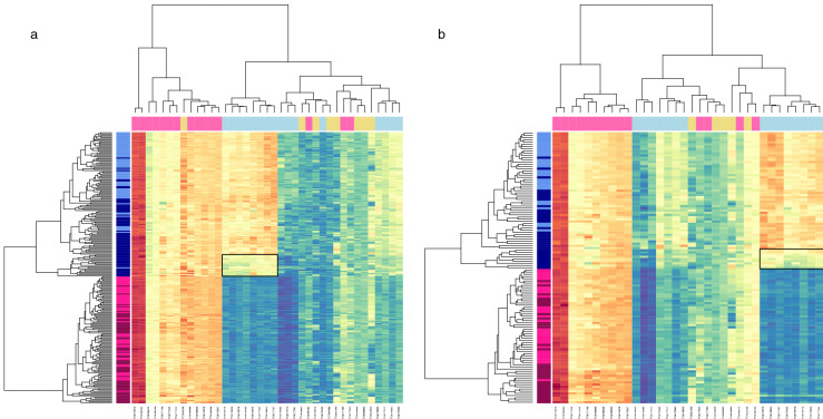
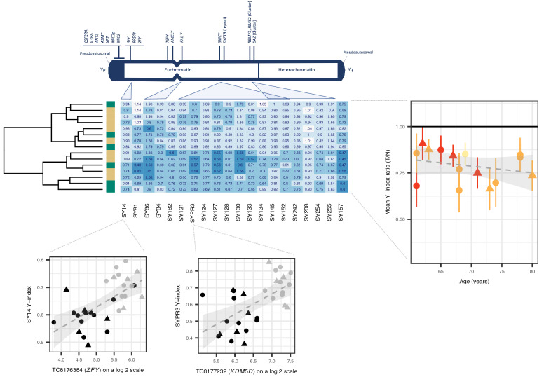
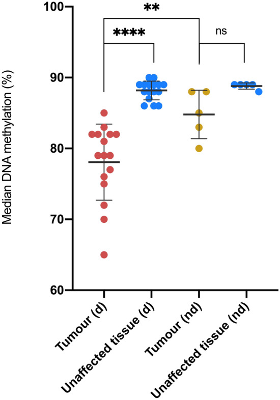
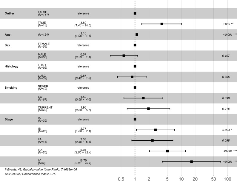
Lung cancer is the most frequent cause of cancer death worldwide. It affects more men than women, and men generally have worse survival outcomes. We compared gene co-expression networks in affected and unaffected lung tissue from 126 consecutive patients with Stage IA-IV lung cancer undergoing surgery with curative intent. We observed marked degradation of a sex-associated transcription network in tumour tissue. This disturbance, detected in 27.7% of male tumours in the discovery dataset and 27.3% of male tumours in a further 123-sample replication dataset, was coincident with partial losses of the Y chromosome and extensive autosomal DNA hypomethylation. Central to this network was the epigenetic modifier and regulator of sexually dimorphic gene expression, KDM5D. After accounting for prognostic and epidemiological covariates including stage and histology, male patients with tumour KDM5D deficiency showed a significantly increased risk of death (Hazard Ratio [HR] 3.80, 95% CI 1.40-10.3, P = 0.009). KDM5D deficiency was confirmed as a negative prognostic indicator in a further 1100 male lung tumours (HR 1.67, 95% CI 1.4-2.0, P = 1.2 × 10⁻¹⁰). Our findings identify tumour deficiency of KDM5D as a prognostic marker and credible mechanism underlying sex disparity in lung cancer.


Conflict of interest statement

A.N. reports personal fees from Merck, Boehringer Ingelheim, Novartis, Astra Zeneca, Bristol Myer Squib, Roche, Abbvie and Oncologica, as well as grants and personal fees from Pfizer outside the submitted work. E.L. reports personal fees from Glaxo Smith Kline, Pfizer, Novartis, Covidien, Roche, Lily Oncology, Boehringer Ingelheim, Medela, Astra Zeneca and Ethicon; Grants and personal fees from ScreenCell; Grants from Clearbridge Biomedics, Illumina and Guardant Health, outside the submitted work. In addition, E.L. has patents P52435GB and P57988GB issued to Imperial Innovations, is the Director of lung screening at the Cromwell Hospital, and is CI for both VIOLET NIHR HTA (13/04/03) and MARS 2 NIHR HTA (15/188/31). S.P. reports personal fees from BMS, Roche, Takeda, AstraZeneca, Pfizer, MSD, EMD, Serono, Guardant Health, Abbvie, Boehringer Ingelheim, OncLive, Medscape, Incyte, Paradox Pharmaceuticals and Eli Lilly outside the submitted work. The other authors declare no competing interests.

Figures

Figure 1
Figure 1
Hierarchical clustering of transcripts assigned to a normal-specific sex associated co-expression network. Figure displays heat maps with hierarchical clustering of samples (on the Y axis) and transcripts (on the X axis) in discovery (a) and replication (b) datasets, limited to transcripts clusters (TC) assigned to the normal-specific, sex associated, gene co-expression network. Expression is shown on a continuous colour scale from blue (low) to red (high). Sample colour (y axis) reflects tissue type (light—histologically normal, dark—tumour) and sex (blue—male, pink—female). Transcript colour (x axis) reflects chromosome class (yellow—autosomal, pink—X, blue—Y). Low Y sample/TCs are highlighted by a solid black box. The data presented in this Figure show broad preservation of a co-expression structure amongst these transcripts in the discovery and replication datasets and confirm the presence of a low Y chromosome expression cluster in a subset of male tumours.
Figure 2
Figure 2
Validation of Loss of Y. Figure is headed with a cartoon adapted from the Promega technical manual depicting sites on chromosome Y interrogated through PCR. A heatmap details the ratios between tumour (T) and histologically normal (N) amplification signals on a patient-by-patient basis. Histology is shown as a y axis sidebar (LUAD = beige, LUSC = green). The grand mean and standard deviation of these ratios across all sites is plotted against age as expressed in years and coloured by smoking history (never smoker = yellow, ex-smoker = orange, current smoker = red). A hatched linear smooth line is shown with its 95% confidence intervals shaded in grey. The relationship between amplification signal (the Y-index, presented on the y axis) and the expression of genes in the same region (presented on the x axis) are shown below, with individual points coloured by tissue class (tumour = black, histologically normal = grey) and including a hatched linear smooth line with 95% confidence intervals shaded in grey. Tumour histology is denoted by point shape (LUAD = circle, LUSC = triangle).
Figure 3
Figure 3
Median CpG DNA methylation percentage per sample. The figure shows median DNA CpG methylation percentage per sample in males with deficient Y chromosome gene expression (d) and males lacking this feature (nd) (see Fig. 1). Data is shown for both tumour and histologically normal tissue. Normality was assessed with a Shapiro Wilk test. Differences in DNA methylation between paired tumour and histologically normal tissues were assessed using a two-tailed paired t-test (low Y group), and a Wilcoxon test (non-low Y group). A two-tailed unpaired Mann–Whitney test was used to assess differences in DNA methylation between the two tumours groups. Error bars represent standard deviation from the mean. Magnitude of significance is denoted with asterisks (*). d deficient chromosome Y gene expression, nd non-deficient chromosome Y gene expression, ns non-significant.
Figure 4
Figure 4
Forest plot for Cox proportional hazards model. The figure provides a forest plot reporting the hazard ratio (HR) and the 95% confidence intervals of the HR for each covariate included in the Cox proportional hazards model. The variable Outlier specifies male tumour samples showing relative KDM5D deficiency (≥ 1.5 SD below the overall male mean). Magnitude of significance is denoted with asterisks (*). LUAD lung adenocarcinoma, LUSC lung squamous cell carcinoma, AIC Akaike information criterion.

References

    1. Cook MB. Epidemiology: Excess cancer in men—A call for an increased research focus. Nat. Rev. Clin. Oncol. 2013;10:186–188. doi: 10.1038/nrclinonc.2013.37. - DOI - PubMed
    1. Cancer Statistics Report . Excess Cancer Burden in Men. Cancer Research; 2013.
    1. Sagerup CM, Smastuen M, Johannesen TB, Helland A, Brustugun OT. Sex-specific trends in lung cancer incidence and survival: A population study of 40,118 cases. Thorax. 2011;66:301–307. doi: 10.1136/thx.2010.151621. - DOI - PubMed
    1. Kinoshita FL, Ito Y, Morishima T, Miyashiro I, Nakayama T. Sex differences in lung cancer survival: Long-term trends using population-based cancer registry data in Osaka, Japan. Jpn. J. Clin. Oncol. 2017;47:863–869. doi: 10.1093/jjco/hyx094. - DOI - PubMed
    1. Wisnivesky JP, Halm EA. Sex differences in lung cancer survival: Do tumors behave differently in elderly women? J. Clin. Oncol. 2007;25:1705–1712. doi: 10.1200/JCO.2006.08.1455. - DOI - PubMed

Publication types

MeSH terms