Review
. 2021 Oct;17(10):639-654.
doi: 10.1038/s41581-021-00430-6. Epub 2021 Jun 14.

Hypertension-induced cognitive impairment: from pathophysiology to public health

Affiliations
Review

Hypertension-induced cognitive impairment: from pathophysiology to public health

Zoltan Ungvari et al. Nat Rev Nephrol. 2021 Oct.

Abstract

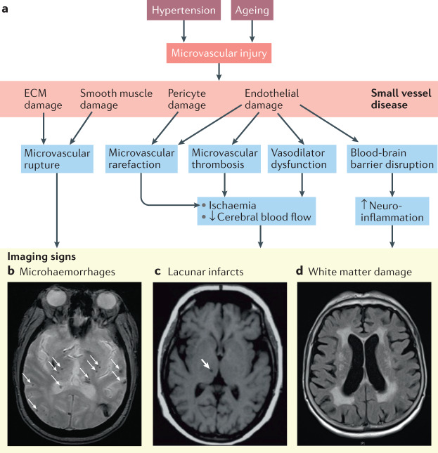
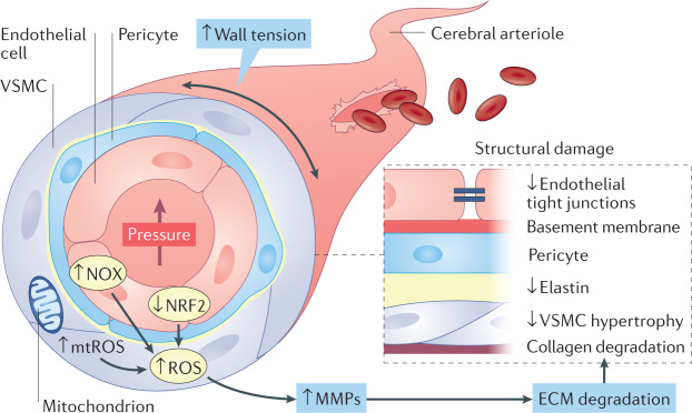
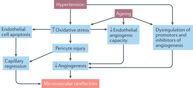
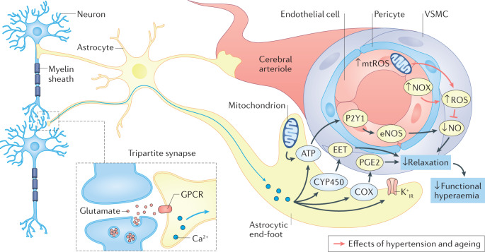
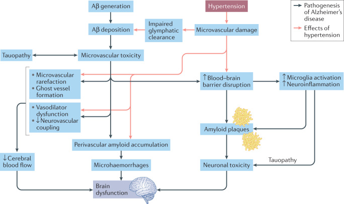
Hypertension affects two-thirds of people aged >60 years and significantly increases the risk of both vascular cognitive impairment and Alzheimer's disease. Hypertension compromises the structural and functional integrity of the cerebral microcirculation, promoting microvascular rarefaction, cerebromicrovascular endothelial dysfunction and neurovascular uncoupling, which impair cerebral blood supply. In addition, hypertension disrupts the blood-brain barrier, promoting neuroinflammation and exacerbation of amyloid pathologies. Ageing is characterized by multifaceted homeostatic dysfunction and impaired cellular stress resilience, which exacerbate the deleterious cerebromicrovascular effects of hypertension. Neuroradiological markers of hypertension-induced cerebral small vessel disease include white matter hyperintensities, lacunar infarcts and microhaemorrhages, all of which are associated with cognitive decline. Use of pharmaceutical and lifestyle interventions that reduce blood pressure, in combination with treatments that promote microvascular health, have the potential to prevent or delay the pathogenesis of vascular cognitive impairment and Alzheimer's disease in patients with hypertension.


Conflict of interest statement

The authors declare no competing interests.

Figures

Fig. 1
Fig. 1. Age-related autoregulatory dysfunction exacerbates hypertension-induced cerebromicrovascular injury.
a | In the vascular smooth muscle cells (VSMCs) of young cerebral arteries, upregulation of a 20-hydroxyeicosatetraenoic-acid (20-HETE)–short transient receptor potential channel 6 (TRPC6)-dependent pathway results in functional adaptation of cerebral arteries to hypertension. Hypertension is associated with increased expression of 20-HETE-producing cytochrome P4504A (CYP4504A) isoforms and TRPC6. High pressure activates phospholipase A2 (PLA2), leading to activation of arachidonic acid (AA) metabolism and the production of 20-HETE, which activates TRPC6 channels, resulting in increases in Ca2+ levels and myogenic contraction. 20-HETE also inhibits activation of hyperpolarizing Ca2+-activated potassium channels (BKca), which facilitates pressure-induced activation of voltage-dependent L-type Ca2+ channels (LCa), contributing to Ca2+ influx and myogenic contraction. This adaptation extends the range of cerebrovascular autoregulatory protection to higher blood pressure levels, optimizing tissue perfusion and protecting the cerebral microcirculation from increased arterial pressure and pressure pulsatility. b | In aged cerebral arteries, functional adaptation to hypertension mediated by activation of the VSMC 20-HETE–TRPC-dependent pathway is impaired. The failure of these arteries to exhibit a hypertension-induced adaptive increase in myogenic constriction results in myogenic contraction and cerebrovascular autoregulatory dysfunction. c | In the aged brain, the failure of proximal resistance arteries to functionally adapt to hypertension results in a mismatch between perfusion pressure and segmental vascular resistance that enables increased pulsatile pressure to penetrate the vulnerable downstream portion of the cerebral microcirculation. The resulting haemodynamic burden exacerbates age-related disruption of the blood–brain barrier (BBB), leading to extravasation of plasma factors (e.g. fibrinogen, thrombin, IgG), which promote microglia activation and neuroinflammation. Microglia-derived pro-inflammatory cytokines, activated matrix metalloproteinases (MMPs) and reactive oxygen species (ROS) induce neuronal damage and synaptic dysfunction,. Increased microvascular pressure impairs the clearance function of the glymphatic (also known as glial-lymphatic) system and promotes the development of cerebral microhaemorrhages via redox-mediated activation of MMPs and consequential weakening of the vascular wall. The increased pressure also contributes to pathological remodelling of the microvascular network architecture by promoting microvascular thrombosis, capillary regression and microvascular rarefaction, resulting in ghost vessels. We posit that exacerbation of neuroinflammation, cerebral microhaemorrhages, glymphatic dysfunction and/or microvascular rarefaction are causally linked to hypertension-induced cognitive impairment in ageing, and also contribute to the pathogenesis of Alzheimer’s disease in hypertensive elderly individuals. Figure adapted with permission from ref., American Physiological Society.
Fig. 2
Fig. 2. Hypertension-induced small vessel disease and its radiological manifestations.
a | Hypertension and ageing promote microvascular injury, including damage to the extracellular matrix (ECM), smooth muscle cells, endothelial cells and pericytes. These effects lead to microvascular rupture, rarefaction and thrombosis as well as impaired vasodilation and blood–brain barrier dysfunction, which result in brain ischaemia and neuroinflammation. This damage is visible as microhaemorrhages, lacunar infarcts and white matter damage on MRI. b | Cerebral microhaemorrhages (arrows) visible on axial T2*-GRE MRI sequences in a 72-year-old man with chronic hypertension, a history of smoking and non-adherence to medical therapy who was admitted for hypertensive emergency with initial blood pressure readings of 230/126 mmHg. The cerebral microhaemorrhages involve the grey–white matter junction and deeper brain regions. c | Silent lacunar infarct (arrow) in the basal ganglia of a 74-year old woman with poorly controlled hypertension who was admitted for confusion. T1-weighted MRI. d | White matter hyperintensities in a 68-year-old man with diabetes mellitus and poorly controlled hypertension who underwent MRI of his head because of progressive worsening of his gait. MRI axial fluid-attenuated inversion recovery sequence image obtained using a 1.5-T field strength scanner.
Fig. 3
Fig. 3. Hypertension-induced blood–brain barrier disruption.
High intraluminal pressure induces increased production of reactive oxygen species (ROS) in the walls of cerebral microvessels. The resulting oxidative stress leads to structural damage to endothelial cells, pericyte injury and increased activation of matrix metalloproteinases (MMPs). Increased MMP activity leads to disruption of tight junctions and breakdown of the extracellular matrix (ECM), resulting in damage to the blood–brain barrier. The damaged blood–brain barrier enables plasma constituents to enter the brain parenchyma, promoting microglia activation, synaptic dysfunction and myelin breakdown. Hypertension-induced neuroinflammation also contributes to synaptic dysfunction and white matter damage. JAM, junctional adhesion molecule; ZO, zonula occludens proteins.
Fig. 4
Fig. 4. Hypertension-induced cerebral microhaemorrhages.
In elderly patients, increased intraluminal pressure and consequential increases in wall tension activate NADPH oxidases (NOX) and promote mitochondria-derived production of reactive oxygen species (mtROS) in the vascular wall. Dysregulation of nuclear factor erythroid 2-related (NRF2)-mediated antioxidant defence mechanisms in the aged vasculature exacerbates pressure-induced oxidative stress. Vascular oxidative stress contributes to increased matrix metalloproteinase (MMP) activation, which promotes degradation of the extracellular matrix (ECM) and vascular smooth muscle cell (VSMC) atrophy. These structural changes weaken the microvascular wall and increase vulnerability to rupture and the formation of cerebral microhaemorrhages. Figure adapted with permission from ref., American Physiological Society.
Fig. 5
Fig. 5. Hypertension and ageing exert synergistic negative effects on cerebromicrovascular network maintenance.
Both hypertension and ageing promote capillary regression and impair angiogenesis. These effects exacerbate cerebromicrovascular rarefaction and compromise cerebral blood supply. The contributing mechanisms include increased oxidative stress-mediated cellular damage and endothelial cell apoptosis, pericyte injury, reduced angiogenic capacity of cerebromicrovascular endothelial cells and dysregulation of promoters and inhibitors of angiogenesis.
Fig. 6
Fig. 6. Hypertension and ageing lead to impairment of endothelium-dependent neurovascular coupling and functional hyperaemia.
Synergistic hypertension-induced and ageing-induced alterations in cerebromicrovascular endothelial cell function and endothelium-dependent neurovascular coupling mechanisms contribute to impaired functional hyperaemia and promote cognitive decline in elderly patients with hypertension. In the healthy brain, a complex interaction between neurons, astrocytes and cerebromicrovascular endothelial cells ensures adequate cerebral blood flow at all times. Neurotransmitters such as glutamate that are released from active excitatory synapses elicit elevations of intracellular Ca2+ concentration in astrocytes via G protein-coupled receptors (GPCRs), initiating the propagation of calcium waves through the processes and soma of the astrocyte to the end-feet, which are wrapped around the resistance arterioles. The surge in astrocyte end-feet Ca2+ concentration promotes ATP release and the cytochrome P450 (CYP450)-mediated and cyclooxygenase (COX)-mediated production of vasodilator eicosanoids (epoxyeicosatrienoic acids (EETs)) and prostaglandins (such as prostaglandin E2 (PGE2)), respectively. Astrocyte-derived ATP promotes endothelial release of the vasodilator nitric oxide (NO) via activation of P2Y purinoceptor 1 (P2Y1). High blood pressure and ageing promote the production of mitochondrial reactive oxygen species (mtROS),, as well as ROS production by NADPH oxidases (NOX),,,. The resulting oxidative stress impairs the bioavailability of endothelial NO and thereby impairs vasodilation, resulting in impairment of functional hyperaemia. Further research is needed to investigate the potential effects of ageing and hypertension on astrocytic regulation of pericyte function and capillary dilation. K+IR, inward rectifier potassium channel; VSMC, vascular smooth muscle cell. Figure adapted with permission from ref., American Physiological Society.
Fig. 7
Fig. 7. Hypertension exacerbates Alzheimer’s disease pathologies.
Alzheimer’s disease is, in part, a microvascular disorder characterized by deposition of the toxic β-amyloid peptide (Aβ) in the brain. This deposition compromises the neurovascular unit and causes multifaceted cerebromicrovascular impairment,. Hypertension may exacerbate the progression of Alzheimer’s disease by exerting synergistic deleterious effects on cells of the neurovascular unit that are already stressed by overproduction of Aβ. Hypertension exacerbates microvascular damage in Alzheimer’s disease and promotes blood–brain barrier disruption and consequential microglia activation, which lead to amyloid plaque formation and neuronal toxicity. In addition, hypertension promotes neurovascular uncoupling and exacerbates capillary atrophy and regression resulting in ghost vessel formation and impaired cerebral blood flow. Perivascular amyloid accumulation facilitated by endothelial damage and Aβ toxicity results in structural damage in arterioles, which promotes the development of microhaemorrhages. Together, these effects contribute to brain dysfunction.

Similar articles

Cited by

References

    1. Hurd MD, Martorell P, Delavande A, Mullen KJ, Langa KM. Monetary costs of dementia in the United States. N. Engl. J. Med. 2013;368:1326–1334. doi: 10.1056/NEJMsa1204629. - DOI - PMC - PubMed
    1. Wimo A, et al. The worldwide economic impact of dementia 2010. Alzheimers Dement. 2013;9:1–11.e3. doi: 10.1016/j.jalz.2012.11.006. - DOI - PubMed
    1. Iadecola C, et al. Impact of hypertension on cognitive function: a scientific statement from the American Heart Association. Hypertension. 2016;68:e67–e94. doi: 10.1161/HYP.0000000000000053. - DOI - PMC - PubMed
    1. Forette F, et al. Prevention of dementia in randomised double-blind placebo-controlled Systolic Hypertension in Europe (Syst-Eur) trial. Lancet. 1998;352:1347–1351. doi: 10.1016/S0140-6736(98)03086-4. - DOI - PubMed
    1. Launer LJ, et al. Midlife blood pressure and dementia: the Honolulu-Asia aging study. Neurobiol. Aging. 2000;21:49–55. doi: 10.1016/S0197-4580(00)00096-8. - DOI - PubMed