

Neuro-ophthalmic manifestations of tuberculosis
- PMID: 34127839
- PMCID: PMC8727585
- DOI: 10.1038/s41433-021-01619-6
Neuro-ophthalmic manifestations of tuberculosis
Abstract
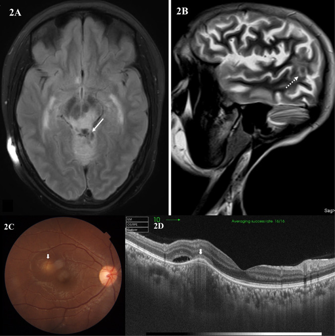
Neuro-ophthalmic features are a known association in tuberculosis, especially common in central nervous system tuberculosis (CNS-TB). They are mostly the result of the visual pathway and/or ocular motor and other cranial nerve involvement. Furthermore, toxic optic neuropathy and paradoxical response to anti-tubercular drugs (ATT) are also not uncommon. The etiopathogenesis is by the complex interplay of various factors like exudates, vasculitis, arachnoiditis, presence of tuberculomas, hydrocephalus, brain infarcts and/or immune-mediated reaction. The entity often poses a diagnostic dilemma for the ophthalmologists/neuro-ophthalmologists and may lead to irreversible vision loss. The presence of neuro-ophthalmic features not only affect the visual outcome but are also predictors of systemic morbidity of the disease. Therefore, understanding and knowledge about this entity are necessary for the comprehensive management of the disease. While various forms of TB including CNS-TB have been well-dealt with in literature, little is discussed specifically about the neuro-ophthalmic manifestations of tuberculosis. Therefore, the purpose of this review is to highlight current understanding of the types of neuro-ophthalmic involvement in tuberculosis, its etiopathogenesis, diagnosis and management.
摘要: 摘要众所周知结核病与神经眼科特征有关, 尤其是在中枢神经系统结核病 (central nervous system tuberculosis, CNS-TB) 常见。它们大多是视觉通路和/或眼运动和其他颅神经受累的结果。此外, 毒性视神经病变和抗结核药物 (anti-tubercular drugs, ATT) 的副作用也并不少见。其病因是由于各种因素如渗出、血管炎、蛛网膜炎、结核瘤、脑积水、脑梗塞和/或免疫因素介导的复杂的相互作用引起的。这种复杂的相互作用网络给眼科医生/神经眼科医生对于疾病诊断的困境, 并可导致不可逆的视力丧失。神经眼科特征的存在不仅影响视力结局, 也是疾病系统发病率的预测因素。因此, 了解和学习这个知识体系对于疾病的综合管理是必要的。虽然文献中对包括CNS-TB在内的各种类型的结核都有很好的论述, 但很少有人针对结核的神经眼科表现进行讨论。因此, 本综述旨在强调结核合并神经眼科疾病的类型、发病机制、诊断和治疗。.
© 2021. The Author(s), under exclusive licence to The Royal College of Ophthalmologists.
Conflict of interest statement
The authors declare no competing interests.
Figures


References
-
- Organization WH. WHO Global tuberculosis report 2019. World Health Organization. 2020. Available from: http://www.who.int/tb/publications/global_report/en/.
-
- WHO. WHO | Tuberculosis and HIV. WHO. 2015. Available from: http://www.who.int/hiv/topics/tb/en/.
-
- Rieder HL, Snider DE, Jr, Cauthen GM. Extrapulmonary tuberculosis in the United States. Am Rev Respir Dis. 1990;141:347–51. - PubMed
-
- CDC. Extrapulmonary tuberculosis cases and percentages by site of disease: reporting areas. Atlanta, GA: Centers Disease Control and Prevention; 2005.
-
- Berger JR. Tuberculous meningitis. Curr Opin Neurol. 1994;7:191–200. - PubMed
Publication types
MeSH terms
LinkOut - more resources
Full Text Sources
Medical
