

Blocking the JAK2/STAT3 and ERK pathways suppresses the proliferation of gastrointestinal cancers by inducing apoptosis
- PMID: 34128372
- PMCID: PMC8214945
- DOI: 10.1631/jzus.B2000842
Blocking the JAK2/STAT3 and ERK pathways suppresses the proliferation of gastrointestinal cancers by inducing apoptosis
Abstract
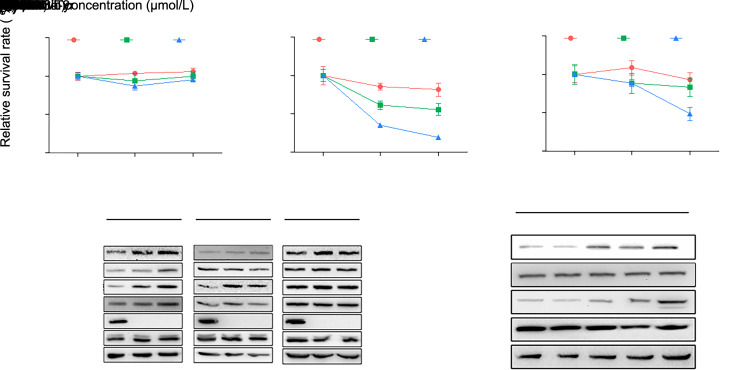
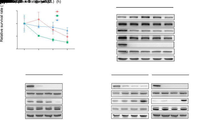
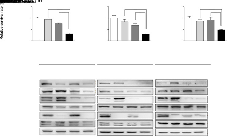
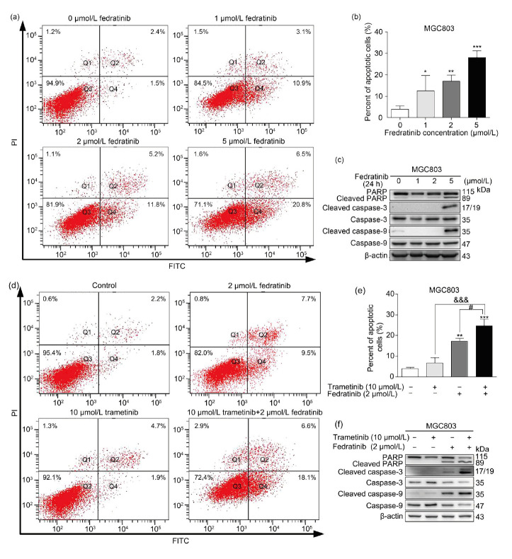
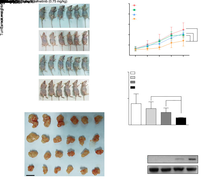
Dysregulated crosstalk between different signaling pathways contributes to tumor development, including resistance to cancer therapy. In the present study, we found that the mitogen-activated extracellular signal-regulated kinase (MEK) inhibitor trametinib failed to suppress the proliferation of PANC-1 and MGC803 cells by activating the Janus kinase 2 (JAK2)/signal transducer and activator of transcription 3 (STAT3) signaling pathway, while the JAK2 inhibitor fedratinib failed to inhibit the growth of the PANC-1 cells upon stimulation of extracellular signal-regulated kinase (ERK) signaling. In particular, the most prominent enhancement of the anti-proliferative effect resulted from the concurrent blockage of the JAK2/STAT3 and ERK signaling pathways. Furthermore, the combination of the two inhibitors resulted in a reduced tumor burden in mice. Our evidence suggests novel crosstalk between JAK2/STAT3 and ERK signaling in gastric cancer (GC) and pancreatic ductal adenocarcinoma (PDAC) cells and provides a therapeutic strategy to overcome potential resistance in gastrointestinal cancer.
Keywords: Apoptosis; Crosstalk; ERK pathway; Gastrointestinal cancers; JAK2/STAT3 pathway.
Figures
Similar articles
-
JAK2 Inhibition Augments the Anti-Proliferation Effects by AKT and MEK Inhibition in Triple-Negative Breast Cancer Cells.Int J Mol Sci. 2025 Jun 26;26(13):6139. doi: 10.3390/ijms26136139. Int J Mol Sci. 2025. PMID: 40649917 Free PMC article.
-
Physapubescin B enhances the sensitivity of gastric cancer cells to trametinib by inhibiting the STAT3 signaling pathway.Toxicol Appl Pharmacol. 2020 Dec 1;408:115273. doi: 10.1016/j.taap.2020.115273. Epub 2020 Oct 6. Toxicol Appl Pharmacol. 2020. PMID: 33035574
-
A new xanthatin analogue 1β-hydroxyl-5α-chloro-8-epi-xanthatin induces apoptosis through ROS-mediated ERK/p38 MAPK activation and JAK2/STAT3 inhibition in human hepatocellular carcinoma.Biochimie. 2018 Sep;152:43-52. doi: 10.1016/j.biochi.2018.06.018. Epub 2018 Jun 28. Biochimie. 2018. PMID: 29960031
-
Unveiling the Role of JAK2/STAT3 signaling in chemoresistance of gynecological cancers: From mechanisms to therapeutic implications.Crit Rev Oncol Hematol. 2025 Jul;211:104712. doi: 10.1016/j.critrevonc.2025.104712. Epub 2025 Apr 3. Crit Rev Oncol Hematol. 2025. PMID: 40187711 Review.
-
Targeting JAK2/STAT3 for the treatment of cancer: A review on recent advancements in molecular development using structural analysis and SAR investigations.Bioorg Chem. 2024 Feb;143:107095. doi: 10.1016/j.bioorg.2023.107095. Epub 2024 Jan 4. Bioorg Chem. 2024. PMID: 38211548 Review.
Cited by
-
Interleukin-6 mediated inflammasome activation promotes oral squamous cell carcinoma progression via JAK2/STAT3/Sox4/NLRP3 signaling pathway.J Exp Clin Cancer Res. 2022 May 5;41(1):166. doi: 10.1186/s13046-022-02376-4. J Exp Clin Cancer Res. 2022. PMID: 35513871 Free PMC article.
-
Typical hemophagocytic syndrome associated with cytomegalovirus infection in an immunocompetent patient: a case report and literature review.J Zhejiang Univ Sci B. 2023 Dec 15;24(12):1159-1164. doi: 10.1631/jzus.B2300232. J Zhejiang Univ Sci B. 2023. PMID: 38057272 Free PMC article. Review.
-
Natural products targeting the MAPK-signaling pathway in cancer: overview.J Cancer Res Clin Oncol. 2024 Jan 9;150(1):6. doi: 10.1007/s00432-023-05572-7. J Cancer Res Clin Oncol. 2024. PMID: 38193944 Free PMC article. Review.
-
Pyroptosis-related genes regulate proliferation and invasion of pancreatic cancer and serve as the prognostic signature for modeling patient survival.Discov Oncol. 2022 May 28;13(1):39. doi: 10.1007/s12672-022-00495-0. Discov Oncol. 2022. PMID: 35633405 Free PMC article.
-
Selenadiazole-Induced Hela Cell Apoptosis through the Redox Oxygen Species-Mediated JAK2/STAT3 Signaling Pathway.ACS Omega. 2024 Apr 30;9(19):20919-20926. doi: 10.1021/acsomega.3c10107. eCollection 2024 May 14. ACS Omega. 2024. PMID: 38764630 Free PMC article.
References
-
- Blair HA, 2019. Fedratinib: first approval. Drugs, 79: 1719-1725https://doi.org/10.1007/s40265- 019-01205-x - PubMed
MeSH terms
Substances
LinkOut - more resources
Full Text Sources
Miscellaneous
