. 2021 Jun 15;131(12):e145800.
doi: 10.1172/JCI145800.

Eruptive xanthoma model reveals endothelial cells internalize and metabolize chylomicrons, leading to extravascular triglyceride accumulation

Affiliations

Eruptive xanthoma model reveals endothelial cells internalize and metabolize chylomicrons, leading to extravascular triglyceride accumulation

Ainara G Cabodevilla et al. J Clin Invest. .

Abstract

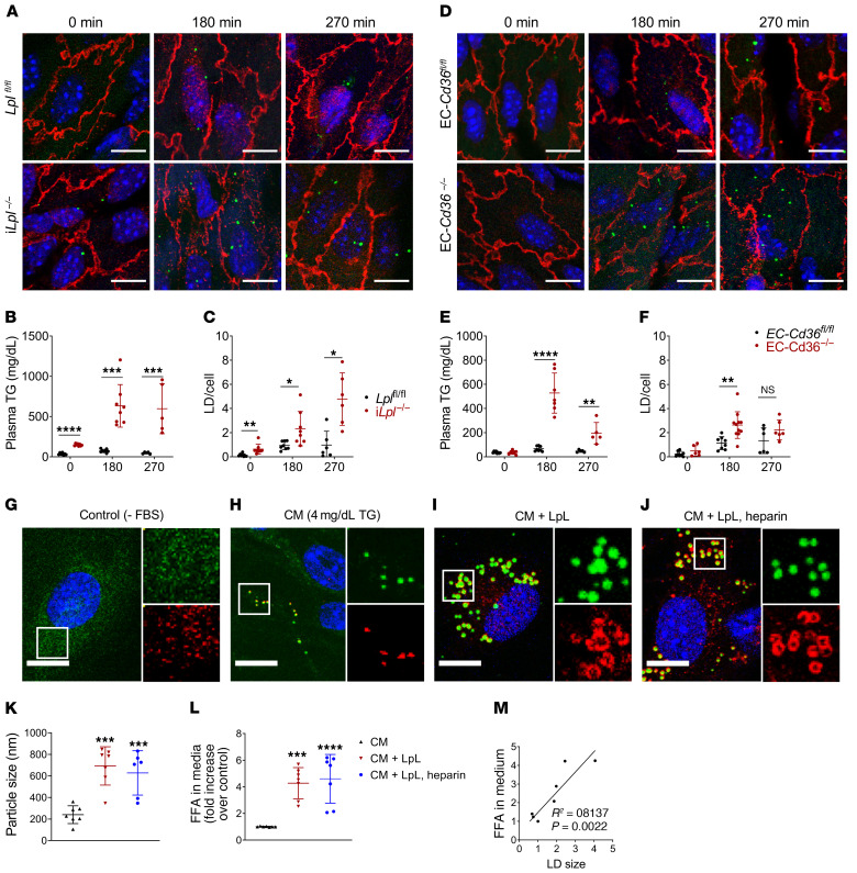
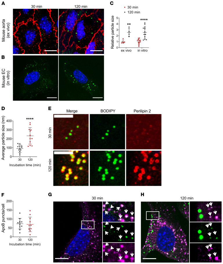
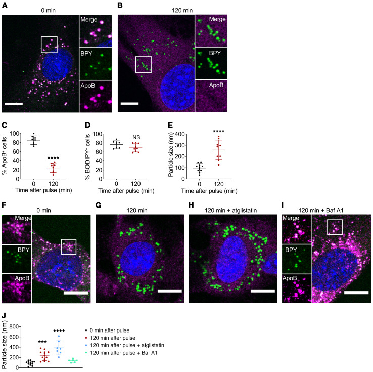
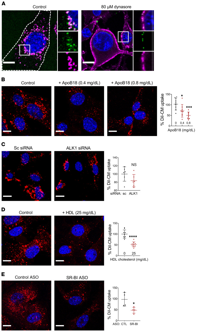
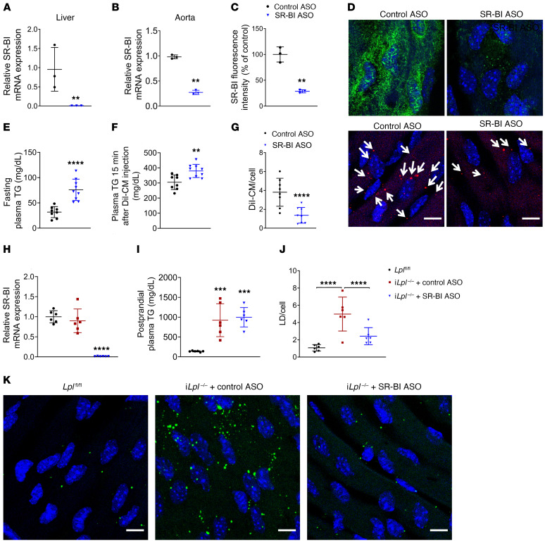
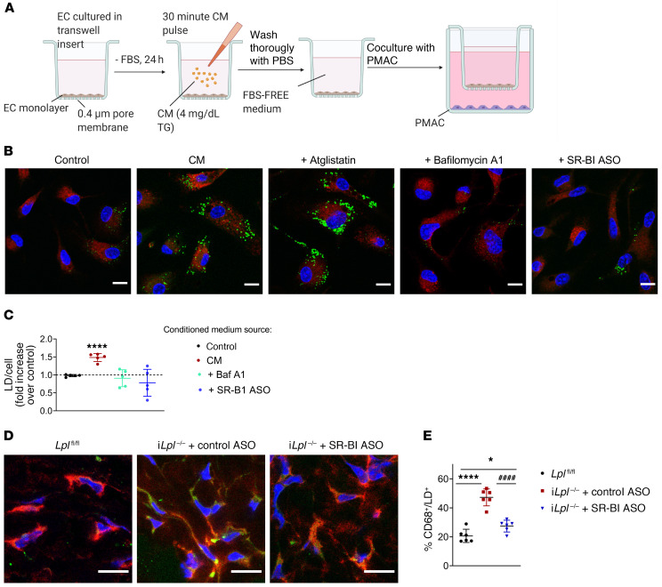
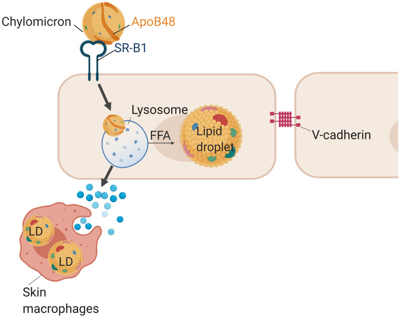
Although tissue uptake of fatty acids from chylomicrons is primarily via lipoprotein lipase (LpL) hydrolysis of triglycerides (TGs), studies of patients with genetic LpL deficiency suggest additional pathways deliver dietary lipids to tissues. Despite an intact endothelial cell (EC) barrier, hyperchylomicronemic patients accumulate chylomicron-derived lipids within skin macrophages, leading to the clinical finding eruptive xanthomas. We explored whether an LpL-independent pathway exists for transfer of circulating lipids across the EC barrier. We found that LpL-deficient mice had a marked increase in aortic EC lipid droplets before and after a fat gavage. Cultured ECs internalized chylomicrons, which were hydrolyzed within lysosomes. The products of this hydrolysis fueled lipid droplet biogenesis in ECs and triggered lipid accumulation in cocultured macrophages. EC chylomicron uptake was inhibited by competition with HDL and knockdown of the scavenger receptor-BI (SR-BI). In vivo, SR-BI knockdown reduced TG accumulation in aortic ECs and skin macrophages of LpL-deficient mice. Thus, ECs internalize chylomicrons, metabolize them in lysosomes, and either store or release their lipids. This latter process may allow accumulation of TGs within skin macrophages and illustrates a pathway that might be responsible for creation of eruptive xanthomas.

Keywords: Endocrinology; Lipoproteins; Metabolism.


Conflict of interest statement

Conflict of interest: AEM is an employee of Ionis Pharmaceuticals.

Figures

Figure 1
Figure 1. Mouse aortic ECs accumulate LDs after an olive oil gavage in the absence of LpL or endothelial CD36.
Three- to four-month-old male iLplfl/fl, iLpl–/–, EC-Cd36fl/fl, and EC-Cd36–/– mice were fasted for 16 hours and given olive oil by oral gavage (10 ml/kg). Plasma TG and aortic EC LD content were assessed at the indicated times (n = 5–8 mice/group/time point). (AC) Both iLplfl/fl and iLpl–/– mice exhibited BODIPY 493/503–positive (green) LDs within aortic ECs, as visualized by confocal microscopy imaging of V-cadherin–immunostained (red) samples (A). Unlike floxed controls, iLpl–/– mice exhibited LDs within aortic ECs prior to the olive oil gavage (A, left panels). In the absence of LpL lipolysis, plasma TG (B) and LD accumulation (C) were significantly increased. (DF) EC-specific CD36 deletion resulted in an increase in aortic EC LD content (D, quantified in F), which was mirrored by elevated plasma TG levels (E). All comparisons are with flox/flox controls. *P < 0.05; **P < 0.01; ***P < 0.001; ****P < 0.0001, 2-way ANOVA. (GM) MECs were deprived of serum overnight to ensure LD depletion (G) and pretreated with LpL (10 U/ml) (I) or LpL+ heparin (10 U/ml). (J) or maintained in FBS-free medium (H) for 1 hour before a 120-minute incubation with human chylomicrons. Samples were immunostained with LD marker perilipin 2 (red) and stained with BODIPY 493/503 (green). LDs were significantly larger in the presence of LpL+/– heparin pretreatment (K), and their size correlated with the presence of FFAs in the media after treatment (M), which was significantly increased in samples pretreated with LpL+/– heparin (L). Data are represented as mean ± SEM of 7 independent experiments. **P < 0.01; ***P < 0.001; ****P < 0.0001, 1-way ANOVA, Dunnet’s multiple comparisons test. Scale bars: 10 μm. Additional inset magnification, ×2.5.
Figure 2
Figure 2. EC BODIPY493/503–positive droplets become larger over time.
(AE) Aortas obtained from fasted WT mice (A) or serum-deprived cultured MECs (B) were incubated with human chylomicrons (4 mg/dL TG in FBS-free medium) for 30 or 120 minutes. Analysis of BODIPY 493/503–positive puncta size (>1000 particles/group) showed a significant increase in particle size over time, with average size increasing more than 2-fold both ex vivo and in vitro (C). Data are represented as mean ± SEM of 4 (ex vivo) or 12 (in vitro) independent experiments. **P < 0.01; ****P < 0.0001, Student’s t test. (D and E) MECs were deprived of serum overnight to ensure LD depletion, then incubated with human chylomicrons (4 mg/dL TG) for the indicated times. (D) Average size of BODIPY 493/503–positive puncta after 30 minutes (~100 nm) or 120 minutes (~250 nm) exposure to chylomicrons. n = 12 independent experiments. ****P < 0.0001, Student’s t test. (E) Immunostaining for perilipin 2 (red). At 120 minutes, BODIPY 493/503–positive droplets (green) colocalized with perilipin 2 (lower panels, colocalization in yellow). There was no perilipin 2 signal or colocalization with BODIPY 493/503–positive droplets at shorter (30 minutes) incubations (upper panels). Scale bars: 5 μm. (FH) Immunostaining for lipoprotein marker apoB. MECs incubated with chylomicrons for 30 or 120 minutes exhibited similar numbers of intracellular apoB-positive puncta (F). At 30-minute incubation, BODIPY 493/503–positive puncta (green) fully colocalized with apoB (magenta, G). Colocalization at 120 minutes was partial, with only smaller BODIPY 493/503–positive puncta colocalizing with apoB (H). White arrows indicate apoB/BODIPY 493/503–positive puncta. Scale bars: 10 μm. Additional inset magnification, ×2.5.
Figure 3
Figure 3. Intracellular chylomicron hydrolysis at the lysosomal compartment precedes LD biogenesis in ECs.
(AE) Cultured MECs were deprived of serum overnight and exposed to human chylomicrons (4 mg/dL in FBS-free medium). After a 30-minute pulse, cells were either fixed (0 minutes) or maintained in FBS-free medium for 120 minutes. (A) As expected, cells fixed immediately after the chylomicron pulse exhibited intracellular apoB/BODIPY 493/503–positive puncta (average size, ~100 nm, E). (B) Incubation for 120 minutes in FBS-free medium after pulse resulted in loss of intracellular apoB signal (quantified in C), accompanied by the appearance of larger, BODIPY 493/503–positive puncta (average size ~ 300 nm, E). Data are represented as mean ± SEM of 8 independent experiments. ****P < 0.0001, Student’s t test. (FI) MECs were pulsed for 30 minutes with human chylomicrons, then fixed (F) or switched for 120 minutes to FBS-free medium alone (G) or in the presence of ATGL inhibitor atglistatin (H) or lysosomal proton pump inhibitor BafA1. (I). Average BODIPY 493/503–positive particle size (n = 7 independent experiments) is represented in J. Inhibition of ATGL did not preclude chylomicron degradation as monitored by loss of intracellular apoB signal nor the appearance of large (~300 nm) LDs. Conversely, inhibition of lysosomal hydrolysis with BafA1 resulted in the retention of apoB/BODIPY 493/503–positivecytoplasmic puncta (~100 nm) for the duration of the treatment. Data are represented as mean ± SD. ***P < 0.001; ****P < 0.0001, 1-way ANOVA, Dunnet’s multiple comparisons test. Scale bars: 10 μm. Additional inset magnification, ×2.
Figure 4
Figure 4. Mechanisms of endothelial chylomicron uptake.
(A) MEC were incubated for 30 minutes with human chylomicrons in the absence (left panel) or presence of the endocytosis inhibitor dynasore (80 μM, right panel). Treatment with dynasore blocked chylomicron uptake, resulting in the accumulation of apoB/BODIPY 493/503–positive chylomicrons in the plasma membrane. Dashed lines show the outline of the cell. (BE) Cultured ECs were deprived of serum overnight, and DiI-labeled human chylomicrons (4 mg/dL TG, red) were used to monitor uptake after a 30-minute pulse. (B) Coincubation with apoB18 peptide (the NH2-terminal sequence of apoB100, 0.4 or 0.8mg/dL) significantly inhibited DiI-chylomicron uptake in MECs. Data are represented as mean ± SD of 8 independent experiments. All comparisons are with control. *P < 0.05; ***P < 0.001, 1-way ANOVA, Dunnet’s multiple comparisons test. (C) Cells treated with control siRNA or ALK1 siRNA were deprived of serum overnight and pulsed for 30 minutes with DiI-labeled chylomicrons. DiI-chylomicron uptake was not significantly reduced in ALK1-deficient cells. (D) DiI-chylomicron uptake was significantly inhibited by coincubation with unlabeled HDL (25 mg/dL cholesterol). (E) MECs were treated with either control or SR-BI ASO for 24 hours, deprived of serum overnight, and pulsed with DiI-chylomicron. Knockdown of SR-BI significantly inhibited DiI-chylomicron uptake. Data are represented as mean ± SEM of 4–9 independent experiments. All comparisons are with control. *P < 0.05; ****P < 0.0001, Student’s t test. Scale bars: 10 μm. Additional inset magnification, ×2.
Figure 5
Figure 5. SR-BI deficiency inhibits in vivo aortic EC CM uptake and LD accumulation.
WT, iLplfl/fl, and iLpl–/– mice (6–8 per group) were injected with either control or SR-BI ASO (100 mg/kg) once a week for 3 weeks. Loss of SR-BI expression was monitored by RT-PCR of liver (A) and aorta (B) as well as by immunostaining of aortic EC with SR-BI (D, green; quantified in C). **P < 0.01, Student’s t test. (EG) DiI-CM (0.5 mg/g TG) was administered retroorbitally to WT mice 72 hours after the last ASO injection. Mice were sacrificed and their aortas harvested 15 minutes after DiI-CM administration, and DiI-CM within aortic ECs were visualized by confocal microscope. Treatment with SR-BI ASO induced fasting hypertriglyceridemia (E), and circulating TG levels were significantly elevated 15 minutes after DiI-CM injection as compared with mice treated with control ASO (F). SR-BI knockdown significantly inhibited DiI-CM uptake in aortic ECs (F, DiI-CM shown in red). White arrows indicate DiI-CM. All comparisons are with control ASO. **P < 0.01; ****P < 0.0001, Student’s t test. (HK) Seventy-two hours after the last ASO injection (H), iLplfl/fl and iLpl–/– mice were fasted overnight and given olive oil by oral gavage (10 ml/kg). Circulating plasma TG levels (I) and aortic EC LD content (J) were assessed 180 minutes after gavage. As expected, iLpl–/– mice treated with control ASO exhibited a significant increase in LD accumulation (K, middle panel) as compared with floxed controls (K, left panel). This phenotype was rescued by SR-BI downregulation (K, right panel). Data are represented as mean ± SD. ***P < 0.001; ****P < 0.0001, 1-way ANOVA, Dunnet’s multiple comparisons test. Scale bars: 10 μm.
Figure 6
Figure 6. SR-BI deficiency reduces LD content in skin macrophages from iLpl–/– mice.
(A) MECs treated with either control or SR-BI ASO were grown to confluency in Transwell inserts and deprived of FBS for 24 hours. On the day of the experiment, ECs were either left untreated (control) or exposed to a 30-minute pulse with chylomicrons, thoroughly washed, and incubated with FBS-free medium with or without atglistatin or BafA1. Inserts were then immediately placed into wells containing freshly harvested PMACs and cocultured for 4 hours. PMACs were immunostained for macrophage marker CD68 (red), and LDs were labeled with BODIPY493/503 (green). (B) Representative images of PMACs following coincubation with ECs treated as indicated. (C) LD/cell quantification. PMACs cocultured with ECs exposed to a chylomicron pulse exhibited significantly more LDs than those cocultured with untreated ECs. LD accumulation was significantly reduced in PMACs cocultured with ECs treated with BafA1 (but not atglistatin) following the chylomicron pulse. SR-BI–deficient ECs exposed to chylomicrons also failed to induce LD biogenesis in cocultured PMACs. Data are represented as mean ± SEM of 5 independent experiments. ****P < 0.0001, 1-way ANOVA. (D and E) iLpl–/– mice were injected with either control or SR-BI ASO. Skin samples from the backs of 6 mice per group, as well as 6 nonhypertriglyceridemic iLplfl/fl controls were immunostained for CD68 (red) and LD staining with BODIPY 493/503 (green). Samples were imaged by confocal microscopy (D), and the percentage of CD68/BODIPY 493/503–positive (yellow) macrophages was quantified (>4000 macrophages per group) (E). LpL deficiency significantly exacerbated LD accumulation in mouse skin macrophages as compared with floxed controls. This effect was significantly reduced in iLpl–/– mice treated with SR-BI ASO. Data are represented as mean ± SD. *P < 0.05; ****P < 0.0001 (significantly different from Lplfl/fl); ####P < 0.0001 (significantly different from iLpl–/–+ control ASO), 1-way ANOVA, Tukey’s multiple comparisons test. Scale bars: 10 μm.
Figure 7
Figure 7. Model.
ECs internalize unhydrolyzed chylomicrons via the SR-BI receptor. Chylomicrons are then intracellularly hydrolyzed within endothelial lysosomes, and the products of this hydrolysis fuel LD biogenesis. Additionally, intracellular chylomicron hydrolysis leads to the release by ECs of biologically active factors capable of triggering LD accumulation in underlying macrophages.

References

    1. Goldberg IJ, Bornfeldt KE. Lipids and the endothelium: bidirectional interactions. Curr Atheroscler Rep. 2013;15(11):365. doi: 10.1007/s11883-013-0365-1. - DOI - PMC - PubMed
    1. Zhang X, et al. Endothelial transcytosis of lipoproteins in atherosclerosis. Front Cardiovasc Med. 2018;5:130. - PMC - PubMed
    1. Goldberg IJ, et al. Regulation of fatty acid uptake into tissues: lipoprotein lipase- and CD36-mediated pathways. J Lipid Res. 2009;50 Suppl(Suppl):S86–S90. - PMC - PubMed
    1. Son NH, et al. Endothelial cell CD36 optimizes tissue fatty acid uptake. J Clin Invest. 2018;128(10):4329–4342. doi: 10.1172/JCI99315. - DOI - PMC - PubMed
    1. Bastie CC, et al. CD36 in myocytes channels fatty acids to a lipase-accessible triglyceride pool that is related to cell lipid and insulin responsiveness. Diabetes. 2004;53(9):2209–2216. doi: 10.2337/diabetes.53.9.2209. - DOI - PubMed

Publication types

MeSH terms