. 2021 Jun 15;131(12):e146051.
doi: 10.1172/JCI146051.

DNA methylome reveals cellular origin of cell-free DNA in spent medium of human preimplantation embryos

Affiliations

DNA methylome reveals cellular origin of cell-free DNA in spent medium of human preimplantation embryos

Yidong Chen et al. J Clin Invest. .

Abstract

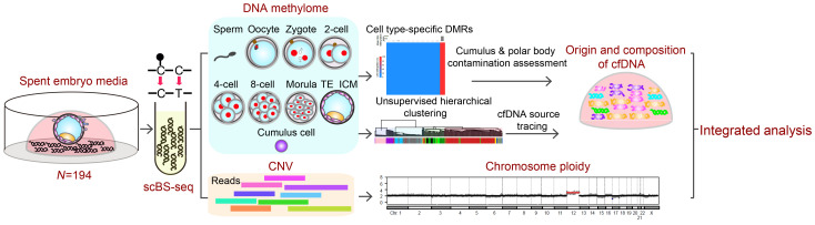
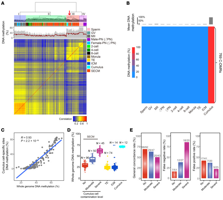
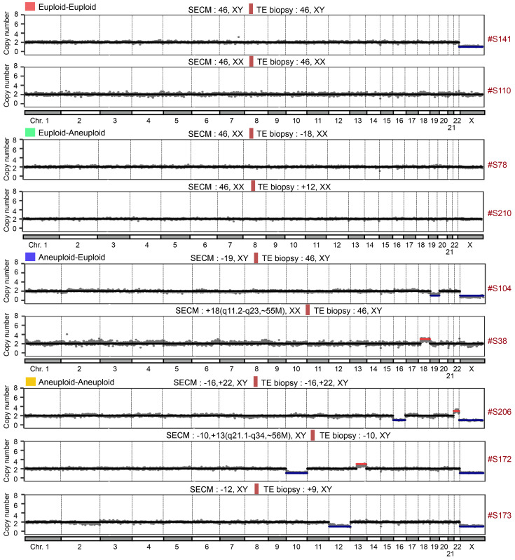
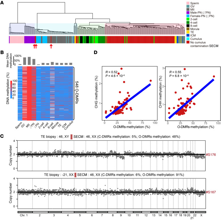
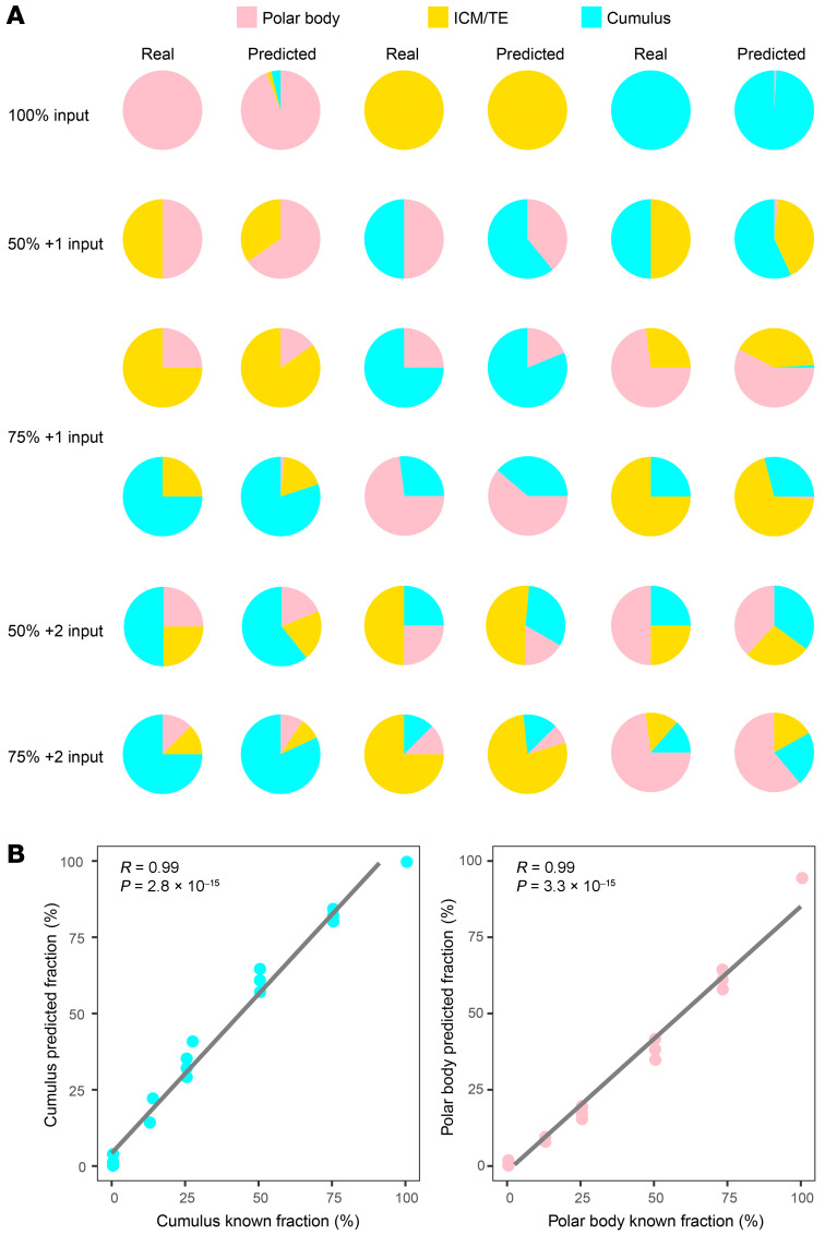
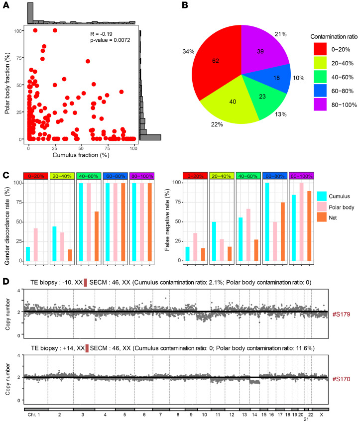
The discovery of embryonic cell-free DNA (cfDNA) in spent embryo culture media (SECM) has brought hope for noninvasive preimplantation genetic testing. However, the cellular origins of SECM cfDNA are not sufficiently understood, and methods for determining maternal DNA contamination are limited. Here, we performed whole-genome DNA methylation sequencing for SECM cfDNA. Our results demonstrated that SECM cfDNA was derived from blastocysts, cumulus cells, and polar bodies. We identified the cumulus-specific differentially methylated regions (DMRs) and oocyte/polar body-specific DMRs, and established an algorithm for deducing the cumulus, polar body, and net maternal DNA contamination ratios in SECM. We showed that DNA methylation sequencing accurately detected chromosome aneuploidy in SECM and distinguished SECM samples with low and high false negative rates and gender discordance rates, after integrating the origin analysis. Our work provides insights into the characterization of embryonic DNA in SECM and provides a perspective for noninvasive preimplantation genetic testing in reproductive medicine.

Keywords: Embryonic development; Epigenetics; Genetics; Molecular diagnosis; Reproductive Biology.


Conflict of interest statement

Conflict of interest: The authors have declared that no conflict of interest exists.

Figures

Figure 1
Figure 1. Study outline.
We performed scBS-seq of SECM, which provided 2 layers of information: DNA methylation and chromosome aneuploidy. We deduced the origin and composition of SECM using the DNA methylome maps of human preimplantation embryos, germ cells, and cumulus cells. scBS-seq was also used to detect chromosome aneuploidy. By calculating the maternal DNA contamination ratio, we can identify the samples with low false negative and gender discordance rates.
Figure 2
Figure 2. Assessment of cumulus contamination in SECM cfDNA.
(A) Unsupervised hierarchical clustering analysis of DNA methylation levels in the SECM cfDNA samples, human preimplantation embryos, germ cells, and cumulus cells. GV, germinal vesicle oocytes; MII, metaphase II oocytes; PN, pronuclei. (B) Heatmap of 769 CpG islands (C-DMRs) that are specifically hypermethylated in cumulus cells. (C) Scatter plot showing a positive correlation between the whole-genome DNA methylation levels and the C-DMR methylation levels in the SECM cfDNAs. The 2-tailed Mann-Whitney-Wilcoxon test was used to assess significance. (D) Box-and-whisker plot showing the whole-genome DNA methylation levels of the ICM, TE, cumulus cells, and 3 SECM cfDNA groups with no, moderate, and severe cumulus contamination degrees as estimated by the C-DMR methylation levels. (E) Bar plots showing the general concordance rate, false negative rate, and false positive rate of the 3 groups of SECM compared with TE biopsy.
Figure 3
Figure 3. scBS-seq detects chromosome aneuploidy in SECM.
Representative CN profiles of SECM in different categories. The results of SECM versus TE biopsy are presented.
Figure 4
Figure 4. Polar body contamination in SECM.
(A) Unsupervised hierarchical clustering of whole-genome DNA methylation for the SECM samples with no cumulus cell contamination, human preimplantation embryos of different stages, germ cells, and cumulus cells. GV, germinal vesicle oocytes; MII, metaphase II oocytes; PN, pronuclei. (B) A total of 548 regions (O-DMRs) were specifically hypermethylated in the MII oocytes. (C) Chromosome CN profiles of 2 SECM samples clustered with the female pronuclei (upper, S167) or the MII oocytes (lower, S176). The chromosome aneuploidy results of TE biopsy and SECM are indicated, along with the methylation levels of the C-DMRs and the O-DMRs. (D) Correlations between non-CpG (left, CHG; right, CHH) DNA methylation levels and the O-DMR DNA methylation levels. CHG/CHH are short for methylation levels on non-CpG islands; H represents A (adenine) or T (thymine). The 2-tailed Mann-Whitney-Wilcoxon test was used to assess significance.
Figure 5
Figure 5. DNA mixing analysis.
(A) Pie charts depicting the results of the simulated DNA mixing experiment. Different percentages of DNA methylation data of the polar body (the MII oocyte), ICM/TE, and cumulus cells were mixed, including 100% input from 1 of the 3 components (100% input), 50% input from each of 2 components (50% + 1 input), 75% input of 1 component plus 25% input of 1 other component (75% + 1 input), 50% input of 1 component plus 25% each of the other 2 components (50% + 2 input), and 75% input of 1 component plus 12.5% each of the other 2 components (75% + 2 input). The input percentages and the predicted percentages are shown for comparison. (B) Correlations between the predicted and input component fractions of the simulated DNA mixing experiment. The 2-tailed Mann-Whitney-Wilcoxon test was used to assess significance.
Figure 6
Figure 6. Maternal DNA ratio in SECM and integrated chromosome aneuploidy analysis.
(A) Scatter plot showing the correlation between the cumulus cell and polar body contamination fractions in SECM. The percentage distribution of each fraction is shown. The 2-tailed Mann-Whitney-Wilcoxon test was used to assess significance. (B) Pie chart showing the numbers and percentages of the SECM samples with different net maternal DNA contamination ratios. (C) Histograms showing GDRs (left) and FNRs (right) for different ratios of cumulus cell, polar body, and net maternal contamination. (D) Representative CN profiles for false negative SECM with nearly no maternal DNA contamination.

Similar articles

Cited by

References

    1. Capalbo A, et al. Correlation between standard blastocyst morphology, euploidy and implantation: an observational study in two centers involving 956 screened blastocysts. Hum Reprod. 2014;29(6):1173–1181. doi: 10.1093/humrep/deu033. - DOI - PubMed
    1. Montag M, et al. Polar body biopsy. Fertil Steril. 2013;100(3):603–607. doi: 10.1016/j.fertnstert.2013.05.053. - DOI - PubMed
    1. Ray PF, et al. Preimplantation genetic diagnosis of beta-thalassaemia major. Lancet. 1996;347(9016):1696. - PubMed
    1. Handyside AH, et al. Biopsy of human preimplantation embryos and sexing by DNA amplification. Lancet. 1989;1(8634):347–349. - PubMed
    1. Kokkali G, et al. Birth of a healthy infant following trophectoderm biopsy from blastocysts for PGD of beta-thalassaemia major. Hum Reprod. 2005;20(7):1855–1859. doi: 10.1093/humrep/deh893. - DOI - PubMed

Publication types