Review
. 2021 Jun 22;144(5):1312-1335.
doi: 10.1093/brain/awab048.

Studying human nociceptors: from fundamentals to clinic

Affiliations
Review

Studying human nociceptors: from fundamentals to clinic

Steven J Middleton et al. Brain. .

Abstract

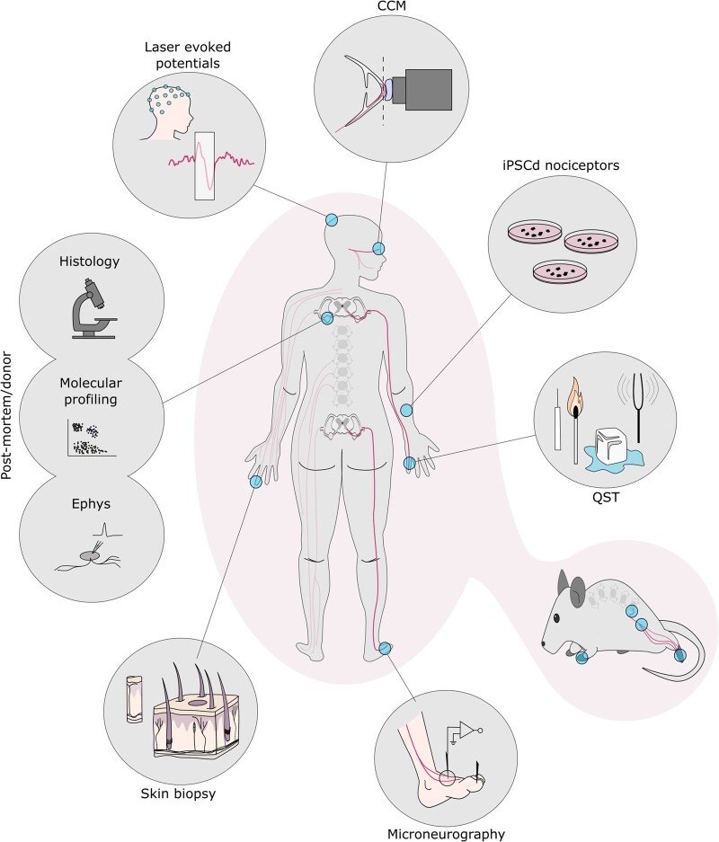
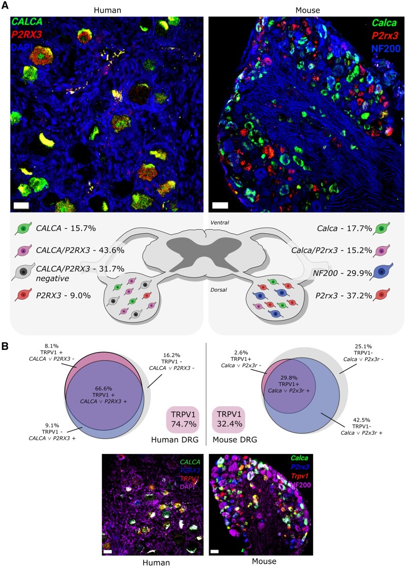
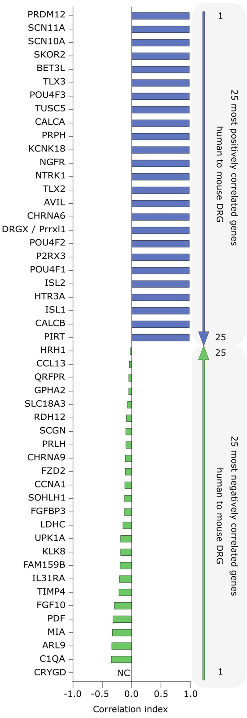
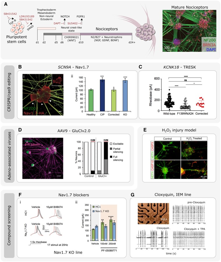
Chronic pain affects one in five of the general population and is the third most important cause of disability-adjusted life-years globally. Unfortunately, treatment remains inadequate due to poor efficacy and tolerability. There has been a failure in translating promising preclinical drug targets into clinic use. This reflects challenges across the whole drug development pathway, from preclinical models to trial design. Nociceptors remain an attractive therapeutic target: their sensitization makes an important contribution to many chronic pain states, they are located outside the blood-brain barrier, and they are relatively specific. The past decade has seen significant advances in the techniques available to study human nociceptors, including: the use of corneal confocal microscopy and biopsy samples to observe nociceptor morphology, the culture of human nociceptors (either from surgical or post-mortem tissue or using human induced pluripotent stem cell derived nociceptors), the application of high throughput technologies such as transcriptomics, the in vitro and in vivo electrophysiological characterization through microneurography, and the correlation with pain percepts provided by quantitative sensory testing. Genome editing in human induced pluripotent stem cell-derived nociceptors enables the interrogation of the causal role of genes in the regulation of nociceptor function. Both human and rodent nociceptors are more heterogeneous at a molecular level than previously appreciated, and while we find that there are broad similarities between human and rodent nociceptors there are also important differences involving ion channel function, expression, and cellular excitability. These technological advances have emphasized the maladaptive plastic changes occurring in human nociceptors following injury that contribute to chronic pain. Studying human nociceptors has revealed new therapeutic targets for the suppression of chronic pain and enhanced repair. Cellular models of human nociceptors have enabled the screening of small molecule and gene therapy approaches on nociceptor function, and in some cases have enabled correlation with clinical outcomes. Undoubtedly, challenges remain. Many of these techniques are difficult to implement at scale, current induced pluripotent stem cell differentiation protocols do not generate the full diversity of nociceptor populations, and we still have a relatively poor understanding of inter-individual variation in nociceptors due to factors such as age, sex, or ethnicity. We hope our ability to directly investigate human nociceptors will not only aid our understanding of the fundamental neurobiology underlying acute and chronic pain but also help bridge the translational gap.

Keywords: IPSC derived nociceptors; microneurography; nociceptors; patch-clamp; transcriptomics.


Figures

Figure 1
Figure 1
Highlighted methods to study human nociceptor anatomy and physiology.Clockwise from top: corneal confocal microscopy (CCM): focal plane (dashed line) lands on the subbasal nerve plexus; IPSC-derived nociceptors; quantitative sensory testing (QST); numerous rodent assays mirror those seen in humans with parallels highlighted throughout this review; microneurography schematic of a peroneal nerve recording; skin biopsy schematic with primary afferents shown in black; post-mortem and donor tissue can be used across applications: histology, molecular profiling and electrophysiology (Ephys); laser evoked potentials, recorded through EEG, measures cortical output in response to heat stimuli.
Figure 2
Figure 2
Classic sensory neuron subpopulations are different between mouse and human DRG. (A) Mouse and human DRG labelled (at ×20) with RNAscope in situ hybridization for CALCA (CGRP; green) and P2RX3 (P2X3R; red) mRNA. Mouse and human DRGs were co-stained for NF200 protein (blue) and DAPI (blue), respectively. Human DRG had a large population of CALCA/P2RX3 co-expressing neurons (43.6%) that was much smaller in mouse (15.2%). This was due to more CALCA being found in human DRG than mouse. These data indicate that the peptidergic (CGRP-positive) and non-peptidergic (P2X3R-positive) neuronal subclasses are not separate entities in human DRG and support that the mouse and human nociceptor phenotypes may be divergent. (B) Mouse and human DRG (at ×20) labelled with RNAscope in situ hybridization for CALCA (CGRP; green), P2RX3 (P2X3R; blue) and TRPV1 (TRPV1; red) mRNA. Mouse and human DRGs were co-stained for NF200 protein (purple) and DAPI (purple), respectively. Trpv1 is expressed in a small percentage of neurons (32.4%) in mouse DRG, most of which are Calca and/or P2rx3 positive (29.8%). However, in human DRG, TRPV1 is expressed in a much larger population (74.7%), the majority of which are positive for CALCA and/or P2RX3. Data shown are summarized from Shiers et al. Scale bar = 50 μm.
Figure 3
Figure 3
Top 25 positively and negatively correlated DRG-enriched gene expression profiles between pharmacologically relevant human and mouse tissues from RNA sequencing. The figure shows the top 25 most positively and negatively correlated (in blue and green respectively) DRG-enriched gene abundance profiles in 12 tissues across human and mouse gene orthologues. The tissues include the DRG, neural tissue (such as spinal cord and brain subregions), and pharmacologically relevant non-neural tissues (such as skeletal muscle and liver). Only genes with relative abundance >0.1 transcripts per kilobase million (TPM) in human DRGs are shown in either list. The methods are described in detail in Ray et al. Among the positively correlated genes, the list is populated with many of the most widely studied genes in the pain and somatosensation fields. Among the anticorrelated genes that are species-specifically enriched in human DRG, most of these genes are relatively unstudied and/or have been validated at the cellular level in independent experiments (e.g. CHRNA9 and IL31RA, discussed in main text). Gene orthologues with different names in human and mice show both names. NC = not calculated since the gene is not expressed in any of the profiled mouse tissues.
Figure 4
Figure 4
Using IPSC-derived nociceptors to develop therapeutics for pain disorders. (A) Illustration of the protocol and timeline of events to generate mature IPSC-derived nociceptors (adapted from Chambers et al. and Meentz et al.; immunomicrograph adapted from Clark et al.121). [B(i)] The use of CRISPR/cas9 gene editing to generate IPSC-derived nociceptors edited to constituently express HA-epitope tagged Nav1.7. (ii) Current-clamp analysis of the current require to elicit and action potential of IPSC-derived nociceptors derived from healthy participants and congenital insensitivity to pain (CIP, due to Nav1.7 loss-of-function) participants. CIP participant IPSC-derived nociceptors were hypoexcitable. The CIP participant hypoexcitability was corrected using CRISPR/Cas9 IPSC editing. CRISPR/Cas9 was used to generate Nav1.7-KO IPSCs that were differentiated into nociceptors that mimicked CIP hypoexcitability (adapted from McDermott et al.106). (C) Current-clamp analysis of the rheobase (minimum current required to elicit and action potential) of IPSC-derived nociceptors from wild-type participants, and participants carrying the F139WfsX24 TRESK loss-of-function mutation. IPSC-derived nociceptors from F139WfsX24 are hyperexcitable compared to wild-type nociceptors. This nociceptor hyperexcitability was rescued when using CRISPR/cas9 to correct the TRESK mutation (adapted from Pettingill et al.103). (D) IPSC-derived nociceptors transduced with the chemogenetic silencing tool GluCl. Scale bar = 50 µm. Patch clamp analysis revealed that following application of the GluCl agonist ivermectin (IVM), GluCl+ nociceptors were rendered either fully silent or partially silent to depolarizing current injections (adapted from Weir et al.122). (E) Example of stem cell derived nociceptors that have been used to model a chemical injury model through the addition of H2O2. H2O2 treatment leads increased caspase expression and neurite retraction/disassembly (adapted from Jones et al.119) [F(i)] IPSC-derived nociceptors derived from healthy control (HC1) or CRIPSR/cas9 engineered Nav1.7 KO lines. The Nav1.7 blocker BIIB074 promoted action potential failure in both HC1 and Nav1.7 KO lines. (ii) Current clamp recordings of HC1 and Nav1.7 KO nociceptors in the presence of vehicle or PF-05089771. Only HC1 but not Nav1.7 KO nociceptors demonstrated changes in excitability in response to the PF-05089771 compound (adapted from McDermott et al.106). (G) Multi-electrode arrays (MEAs) used to assess spontaneous activity of IPSC-derived nociceptors from an IEM participant. Activity at electrodes was reduced after addition of the TRESK activator cloxyquin. Spontaneous activity recovery was lost following co-addition of TPA (TRESK inhibitor) and cloxyquin (adapted from Pettingill et al.103).
Figure 5
Figure 5
Properties of human versus rat DRG neurons and their response to selective blockers in vitro. (A) The representative immunohistochemistry illustrates that Nav1.7, Cav3.2 and TRPV1 are expressed in naïve rat DRG (iiii) and this expression is elevated in rats with neuropathic pain produced using paclitaxel treatment (ivvi). All three channels are also expressed at relatively low levels in human DRG where neuropathic pain is absent (viiix), but also become markedly elevated when neuropathic pain is present (xxii). (B) The representative analogue traces in iiv show four common types of action potential waveforms observed in whole cell recordings from human DRG neurons. The representative analogue recordings in vvii show the three typical patterns of response shown by human DRG neurons to intracellular currents using graded steps in intensity from rheobase as colour coded at top right. (C) The representative analogue recordings in show that the Nav1.7 inhibitor ProTxII, given at the time indicated by the solid line over each trace, inhibits spontaneous activity in both rat (i) and human (ii) neurons. Note the downward (hyperpolarizing) shift in the resting membrane potential in the rat DRG neuron recording during ProTxII administration. (D) The representative analogue recordings in show that the Cav3.2 inhibitor ML218, given at the time indicated by the solid line over each trace, inhibits spontaneous activity in both rat (i) and human (ii) neurons. Note the downward (hyperpolarizing) shift in the resting membrane potential in the human DRG neuron recording during ML218 administration.

References

    1. Sherrington CS. The integrative action of the nervous system. C. Scribner's sons; 1906.
    1. Chisholm KI, Khovanov N, Lopes DM, La Russa F, McMahon SB.. Large scale in vivo recording of sensory neuron activity with GCaMP6. eNeuro. 2018;5(1):1–14. ENEURO.0417-17.2018. - PMC - PubMed
    1. Emery EC, Luiz AP, Sikandar S, Magnúsdóttir R, Dong X, Wood JN.. In vivo characterization of distinct modality-specific subsets of somatosensory neurons using GCaMP. Sci Adv. 2016;2(11):e1600990. - PMC - PubMed
    1. Lawson SN, Fang X, Djouhri L.. Nociceptor subtypes and their incidence in rat lumbar dorsal root ganglia (DRGs): Focussing on C-polymodal nociceptors, Abeta-nociceptors, moderate pressure receptors and their receptive field depths. Curr Opin Physiol. 2019;11:125–146. - PMC - PubMed
    1. Schmidt R, Schmelz M, Forster C, Ringkamp M, Torebjork E, Handwerker H.. Novel classes of responsive and unresponsive C nociceptors in human skin. J Neurosci. 1995;15(1):333–341. - PMC - PubMed

Publication types