

Selective 13 C-Labels on Repeating Glycan Oligomers to Reveal Protein Binding Epitopes through NMR: Polylactosamine Binding to Galectins
- PMID: 34128568
- PMCID: PMC8456918
- DOI: 10.1002/anie.202106056
Selective 13 C-Labels on Repeating Glycan Oligomers to Reveal Protein Binding Epitopes through NMR: Polylactosamine Binding to Galectins
Abstract
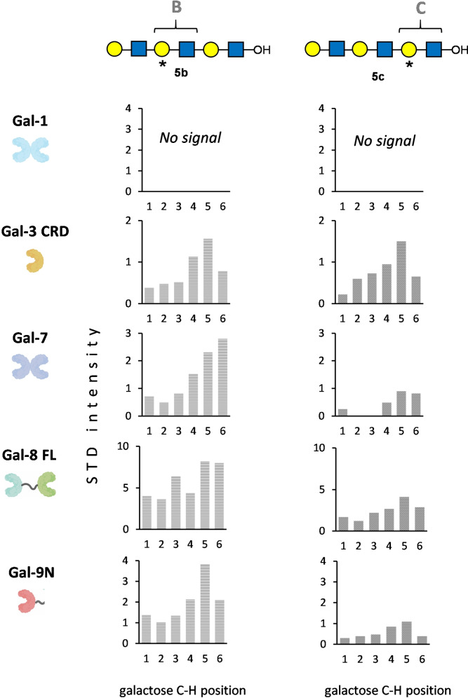
A combined chemo-enzymatic synthesis/NMR-based methodology is presented to identify, in unambiguous manner, the distinctive binding epitope within repeating sugar oligomers when binding to protein receptors. The concept is based on the incorporation of 13 C-labels at specific monosaccharide units, selected within a repeating glycan oligomeric structure. No new chemical tags are added, and thus the chemical entity remains the same, while the presence of the 13 C-labeled monosaccharide breaks the NMR chemical shift degeneracy that occurs in the non-labeled compound and allows the unique identification of the different components of the oligomer. The approach is demonstrated by a proof-of-concept study dealing with the interaction of a polylactosamine hexasaccharide with five different galectins that display distinct preferences for these entities.
Keywords: NMR; galectins; molecular recognition; polylactosamine; selective 13C-labels.
© 2021 The Authors. Angewandte Chemie International Edition published by Wiley-VCH GmbH.
Conflict of interest statement
The authors declare no conflict of interest.
Figures


Similar articles
-
NMR Investigation of Protein-Carbohydrate Interactions: The Recognition of Glycans by Galectins Engineered with Fluorotryptophan Residues.Chemistry. 2023 Jan 24;29(5):e202202208. doi: 10.1002/chem.202202208. Epub 2022 Dec 12. Chemistry. 2023. PMID: 36343278 Free PMC article.
-
Exploring Glycan-Lectin Interactions in Natural-Like Environments: A View Using NMR Experiments Inside Cell and on Cell Surface.Chemistry. 2025 Feb 17;31(10):e202403102. doi: 10.1002/chem.202403102. Epub 2024 Dec 11. Chemistry. 2025. PMID: 39588609 Free PMC article.
-
Comparative study of the glycan specificities of cell-bound human tandem-repeat-type galectin-4, -8 and -9.Glycobiology. 2012 Sep;22(9):1207-17. doi: 10.1093/glycob/cws079. Epub 2012 Apr 30. Glycobiology. 2012. PMID: 22547138
-
Specificity of human galectins on cell surfaces.Biochemistry (Mosc). 2015 Jul;80(7):846-56. doi: 10.1134/S0006297915070056. Biochemistry (Mosc). 2015. PMID: 26541999 Review.
-
Galactoseβ1-4fucose: A unique disaccharide unit found in N-glycans of invertebrates including nematodes.Proteomics. 2016 Dec;16(24):3137-3147. doi: 10.1002/pmic.201600001. Epub 2016 Jun 8. Proteomics. 2016. PMID: 27091793 Review.
Cited by
-
Exploring multivalent carbohydrate-protein interactions by NMR.Chem Soc Rev. 2023 Mar 6;52(5):1591-1613. doi: 10.1039/d2cs00983h. Chem Soc Rev. 2023. PMID: 36753338 Free PMC article. Review.
-
Decoding Strategies to Evade Immunoregulators Galectin-1, -3, and -9 and Their Ligands as Novel Therapeutics in Cancer Immunotherapy.Int J Mol Sci. 2022 Dec 8;23(24):15554. doi: 10.3390/ijms232415554. Int J Mol Sci. 2022. PMID: 36555198 Free PMC article. Review.
-
Analysis of carbohydrates and glycoconjugates by matrix-assisted laser desorption/ionization mass spectrometry: An update for 2021-2022.Mass Spectrom Rev. 2025 May-Jun;44(3):213-453. doi: 10.1002/mas.21873. Epub 2024 Jun 24. Mass Spectrom Rev. 2025. PMID: 38925550 Free PMC article. Review.
-
NMR Investigation of Protein-Carbohydrate Interactions: The Recognition of Glycans by Galectins Engineered with Fluorotryptophan Residues.Chemistry. 2023 Jan 24;29(5):e202202208. doi: 10.1002/chem.202202208. Epub 2022 Dec 12. Chemistry. 2023. PMID: 36343278 Free PMC article.
-
Exploring Glycan-Lectin Interactions in Natural-Like Environments: A View Using NMR Experiments Inside Cell and on Cell Surface.Chemistry. 2025 Feb 17;31(10):e202403102. doi: 10.1002/chem.202403102. Epub 2024 Dec 11. Chemistry. 2025. PMID: 39588609 Free PMC article.
References
-
- None
-
- Zhang Y., Gómez-Redondo M., Jiménez-Osés G., Ardá A., Overkleeft H. S., Marel G. A., Jiménez-Barbero J., Codée J. D. C., Angew. Chem. Int. Ed. 2020, 59, 12746–12750; - PubMed
- Angew. Chem. 2020, 132, 12846–12850;
-
- Gómez-Redondo M., Ardá A., Gimeno A., Jiménez-Barbero J., Drug Discovery Today Technol. 2020, 35–36, 1–11; - PubMed
Publication types
MeSH terms
Substances
LinkOut - more resources
Full Text Sources
