Review
. 2021 Jun;11(6):210137.
doi: 10.1098/rsob.210137. Epub 2021 Jun 16.

Higher-order organization of biomolecular condensates

Affiliations
Review

Higher-order organization of biomolecular condensates

Charlotte M Fare et al. Open Biol. 2021 Jun.

Abstract

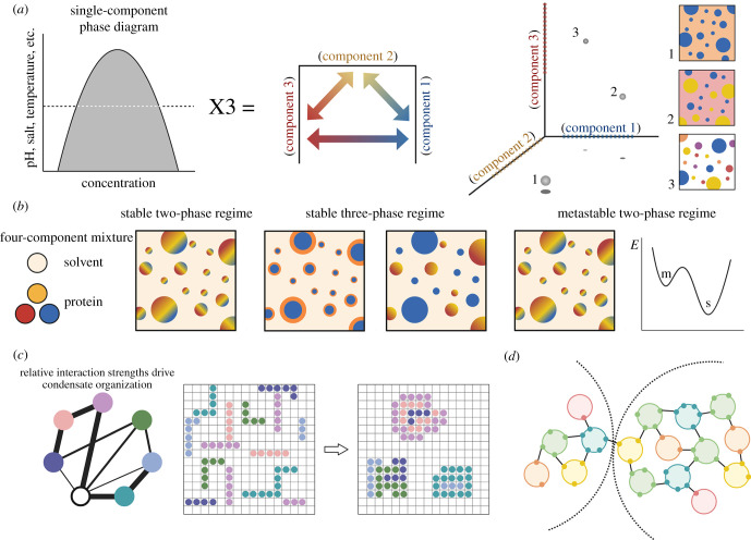
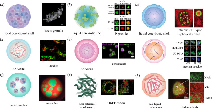
A guiding principle of biology is that biochemical reactions must be organized in space and time. One way this spatio-temporal organization is achieved is through liquid-liquid phase separation (LLPS), which generates biomolecular condensates. These condensates are dynamic and reactive, and often contain a complex mixture of proteins and nucleic acids. In this review, we discuss how underlying physical and chemical processes generate internal condensate architectures. We then outline the diverse condensate architectures that are observed in biological systems. Finally, we discuss how specific condensate organization is critical for specific biological functions.

Keywords: biomolecular condensate; liquid–liquid phase separation; membraneless organelle.


Figures

Figure 1.
Figure 1.
Physical and chemical principles underlie the internal organization adopted by biomolecular condensates. (a) In a classic single-component phase diagram (left), the x-axis represents protein concentration, and the y-axis represents another variable, such as temperature or salt concentration. However, in biological systems, proteins are not typically in isolation. Therefore, the phase behaviour of any given protein is affected by the properties and abundance of the other proteins in its environment. A simplified schema to consider the interplay between three different protein components in a given environment (represented on each axis, where the dashed line on each axis represents the concentration at which a given component will phase separate within a shared environment) is modelled on the right. As illustrated, variations in the concentration of each protein affect which proteins are condensed, whereas other chemical factors influence whether two protein condensates are miscible. In condition 1, component 1 (blue) is condensed, and components 2 (yellow) and 3 (red) are mixed in the light phase (orange). In condition 2, component 3 remains in the light phase, whereas components 1 and 2 form independent condensates. In condition 3, all components are condensed; in addition to each component forming a homogeneous condensate, components 1 and 2 can each form a heterogeneous condensate with component 3 (purple and orange, respectively). (b) In addition to coexisting condensed phases, complex mixtures can also form ordered condensates. For example, in a four-component mixture in which two components repel each other, there are three potential regimes. The first is a stable two-phase state in which three of the four components form miscible condensates. The second state is a stable three-phase regime, in which two of the three condensed components are miscible with each other, whereas one is separate. The characteristics of this state depend on the relative concentrations of the components and their interactions with one another. The third regime that can arise from a four-component mixture is a metastable state, which superficially resembles the stable two-phase state. However, a metastable state can only exist in the absence of energetic noise. On a free-energy diagram, this state exists at a local energy minimum (m), but with thermal perturbations, the mixture will shift towards a more stable state (s). (c) To understand how condensates form, many researchers have used computational approaches, including lattice modelling [24]. In a lattice model, proteins are represented as polypeptide chains, and the interactions between proteins and with solvent can be parameterized within the simulation. Then, protein chains will self-associate or segregate based on these chemical attributes. Lattice modelling can be useful as both a descriptive and predictive tool for studying biomolecular condensate behaviour. (d) One way to conceptualize condensates is as a network of particles in which each particle has some valency [25]. In such a network, components can be classified based on their valency, where a valency of one prevents further network growth, whereas bridges and nodes (capable of forming 2 or 3+ interactions, respectively) promote condensate expansion and association. In this framework, a condensate only forms when there are sufficient cross-links between network particles. The extent to which two condensates interact can be determined by identifying shared nodes between networks. This figure was made with BioRender.
Figure 2.
Figure 2.
Examples of biomolecular condensate architecture. (a) Solid core-liquid shell. Condensates with this structure initially form as individual protein cores, which then accumulate more material and join together to form larger structures, as with SGs. Green dots indicate G3BP cores and grey represents the surface area of the SG; scale bar represents 500 nm [83]. (b) Liquid core-solid shell. Condensates with this structure consist of a more liquid-like core surrounded by a gel-like protein shell. An example of this architecture is the P granule found in C. elegans embryos. Condensates formed by MEG-3 (green) and PGL-3 (red) in cells and in vitro show this architecture; scale bars represent 1 µm [84]. (c) Liquid core-liquid shell. With this architecture, the interior and the exterior of the condensate are both dynamic liquids, but are compositionally distinct, such as with iLSA. iLSA have a liquid TDP-43 shell (green) and the interior is enriched for HSP70 chaperones, including Hsc70 (red); scale bar represents 5 µm [85]. (d) RNA core. In X. laevis oocytes, RNAs must properly localize for successful embryonic development. This localization is achieved, in part, by the formation of L-bodies, composed of RNAs and RBPs. In L-bodies, the RNA forms an immobile core enveloped by a dynamic protein coating. Staining for the L-body protein hnRNPAB (green) and mRNA vg1 (red) reveals the multiphasic nature of these condensates; scale bar represents 10 µm [86]. (e) RNA shell. There are multiple ways in which a condensate can form such that protein is surrounded by RNA. In paraspeckles (left), the lncRNA NEAT1 folds so that its 3′ and 5′ ends face outward with its middle at the centre where proteins accumulate. Labelling the 5′ and 3′ ends of NEAT1 (green) highlights the paraspeckle shell, whereas labelling the middle portion of NEAT1 or a protein such as Nono (purple) identifies the core; scale bar represents 500 nm [87]. Alternatively, RNAs can decorate the exterior of a protein core, as with nuclear speckles (right). In this situation, a proteinaceous core collects RNA molecules that reside on the outside of the condensate. SIM images of nuclear speckles show splicing protein SC35 (blue) occupying a central area, with lncRNA MALAT1 (red) and U2 RNA (green) taking up an area with a larger radius; scale bars represent 1 µm [33]. (f) Nested Droplets. The nucleolus is an example of a condensate with a nested droplet architecture. The nucleolus has a tripartite organization, with the gel-like FC and DFC enveloped by a liquid-like GC. Each component of the nucleolus has a distinct function and displays distinct physical properties. In X. laevis nuclei, RNA Pol I (blue) localizes to the innermost FC, FIB1 (green) stains the DFC, and NPM1 (red) is localized at the liquid-like GC; scale bar represents 20 µm [88]. (g) Non-spherical condensates. Biomolecular condensates can also adopt atypical structures, as in the case of TIS11B granules. TIS11B granules form an extensive mesh-like assembly composed of protein and RNA. In cells, TIS11B condensates (red) intertwine with the ER (green), forming a complex network of tubules; scale bars indicate 5 µm (left) and 1 µm (right) [89]. (h) Non-liquid condensates. In addition to liquid-like condensates, biomolecules can also condense to form solid states. One example of this is the Balbiani body, which contains an amyloidogenic network formed by the protein Xvelo in X. laevis (homologous to the Bucky ball protein in zebrafish). Xvelo (green) contains a PrLD which is necessary for its self-assembly, as well as its association with organelles, like mitochondria (red); scale bars indicate 20 µm and 2 µm (insets) [90]. This figure was made with BioRender.

References

    1. Flamholz A, Phillips R, Milo R. 2014. The quantified cell. MBoC 25, 3497-3500. ( 10.1091/mbc.e14-09-1347) - DOI - PMC - PubMed
    1. Shorter J, Warren G. 2002. Golgi architecture and inheritance. Annu. Rev. Cell Dev. Biol. 18, 379-420. ( 10.1146/annurev.cellbio.18.030602.133733) - DOI - PubMed
    1. Hyman AA, Weber CA, Jülicher F. 2014. Liquid-liquid phase separation in biology. Annu. Rev. Cell Dev. Biol. 30, 39-58. ( 10.1146/annurev-cellbio-100913-013325) - DOI - PubMed
    1. Mitrea DM, Kriwacki RW. 2016. Phase separation in biology; functional organization of a higher order. Cell Commun. Signal 14, 1. ( 10.1186/s12964-015-0125-7) - DOI - PMC - PubMed
    1. Boeynaems S, et al. 2018. Protein phase separation: a new phase in cell biology. Trends Cell Biol. 28, 420-435. ( 10.1016/j.tcb.2018.02.004) - DOI - PMC - PubMed

Publication types