

Artificial Intelligence and Cellular Segmentation in Tissue Microscopy Images
- PMID: 34129842
- PMCID: PMC8485056
- DOI: 10.1016/j.ajpath.2021.05.022
Artificial Intelligence and Cellular Segmentation in Tissue Microscopy Images
Abstract
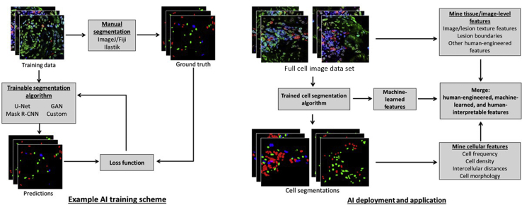
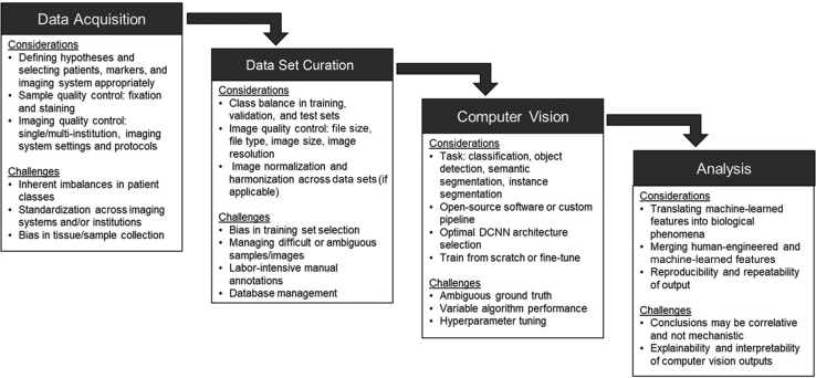
With applications in object detection, image feature extraction, image classification, and image segmentation, artificial intelligence is facilitating high-throughput analysis of image data in a variety of biomedical imaging disciplines, ranging from radiology and pathology to cancer biology and immunology. Specifically, a growth in research on deep learning has led to the widespread application of computer-visualization techniques for analyzing and mining data from biomedical images. The availability of open-source software packages and the development of novel, trainable deep neural network architectures has led to increased accuracy in cell detection and segmentation algorithms. By automating cell segmentation, it is now possible to mine quantifiable cellular and spatio-cellular features from microscopy images, providing insight into the organization of cells in various pathologies. This mini-review provides an overview of the current state of the art in deep learning- and artificial intelligence-based methods of segmentation and data mining of cells in microscopy images of tissue.
Copyright © 2021 American Society for Investigative Pathology. Published by Elsevier Inc. All rights reserved.
Figures
References
-
- Wang H., Shang S., Long L., Hu R., Wu Y., Chen N., Zhang S., Cong F., Lin S. Biological image analysis using deep learning-based methods: literature review. Digit Med. 2018;4:157–165.
Publication types
MeSH terms
Grants and funding
LinkOut - more resources
Full Text Sources
Other Literature Sources
