. 2021 Oct;75(4):912-923.
doi: 10.1016/j.jhep.2021.05.041. Epub 2021 Jun 12.

Loss of hepatocyte identity following aberrant YAP activation: A key mechanism in alcoholic hepatitis

Affiliations

Loss of hepatocyte identity following aberrant YAP activation: A key mechanism in alcoholic hepatitis

Mohamed Bou Saleh et al. J Hepatol. 2021 Oct.

Abstract

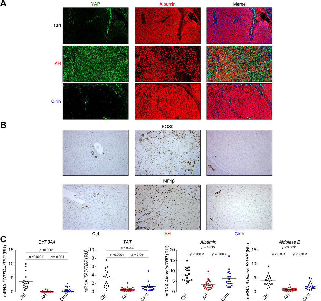
Background & aims: Alcoholic hepatitis (AH) is a life-threatening disease with limited therapeutic options, as the molecular mechanisms leading to death are not well understood. This study evaluates the Hippo/Yes-associated protein (YAP) pathway which has been shown to play a role in liver regeneration.

Method: The Hippo/YAP pathway was dissected in explants of patients transplanted for AH or alcohol-related cirrhosis and in control livers, using RNA-seq, real-time PCR, western blot, immunohistochemistry and transcriptome analysis after laser microdissection. We transfected primary human hepatocytes with constitutively active YAP (YAPS127A) and treated HepaRG cells and primary hepatocytes isolated from AH livers with a YAP inhibitor. We also used mouse models of ethanol exposure (Lieber de Carli) and liver regeneration (carbon tetrachloride) after hepatocyte transduction of YAPS127A.

Results: In AH samples, RNA-seq analysis and immunohistochemistry of total liver and microdissected hepatocytes revealed marked downregulation of the Hippo pathway, demonstrated by lower levels of active MST1 kinase and abnormal activation of YAP in hepatocytes. Overactivation of YAP in hepatocytes in vitro and in vivo led to biliary differentiation and loss of key biological functions such as regeneration capacity. Conversely, a YAP inhibitor restored the mature hepatocyte phenotype in abnormal hepatocytes taken from patients with AH. In ethanol-fed mice, YAP activation using YAPS127A resulted in a loss of hepatocyte differentiation. Hepatocyte proliferation was hampered by YAPS127A after carbon tetrachloride intoxication.

Conclusion: Aberrant activation of YAP plays an important role in hepatocyte transdifferentiation in AH, through a loss of hepatocyte identity and impaired regeneration. Thus, targeting YAP is a promising strategy for the treatment of patients with AH.

Lay summary: Alcoholic hepatitis is characterized by inflammation and a life-threatening alteration of liver regeneration, although the mechanisms behind this have not been identified. Herein, we show that liver samples from patients with alcoholic hepatitis are characterized by profound deregulation of the Hippo/YAP pathway with uncontrolled activation of YAP in hepatocytes. We used human cell and mouse models to show that inhibition of YAP reverts this hepatocyte defect and could be a novel therapeutic strategy for alcoholic hepatitis.

Keywords: Hippo/YAP; alcoholic hepatitis; hepatocyte; regeneration; transdifferentiation.


Conflict of interest statement

Conflict of interest The authors declare that they have no competing interests in relation to this manuscript. Please refer to the accompanying ICMJE disclosure forms for further details.

Figures

Fig. 1.
Fig. 1.. Abnormal YAP activation in liver samples from patients with AH.
(A) A heat map showing mRNA expression levels of Hippo/YAP pathway components and their partners in AH liver explants (exAH, in purple), liver biopsies from patients with severe AH (red), liver biopsies from patients with early-stage AH (eASH, in blue) and liver biopsies from patients with normal livers (N, in green). The fold change is the relative variation of gene expression to the center value (mean of all values of that gene in the population) (B) Real-time PCRs performed on liver samples from Ctrl patients, patients with AH and patients with Cirrh. mRNA expression levels of YAP1, its target genes (NUAK2, ANKRD1, and TGFβ2) and its cofactor TEAD4 were compared with the TBP housekeeping gene (coding for TATA-binding protein). (C) A representative western blot of active and inactive MST1 in the liver. Beta-actin was used as a loading control. The dot plots illustrate the band intensity for total MST1 and active MST1. The results are expressed as total MST1/β-actin and active MST1/β-actin ratios, in relative units. (D) Representative YAP immunostaining experiments in livers from Ctrl, AH and Cirrh groups at a magnification of x100 (upper panel) or x200 (lower panel). In control livers, the portal bile ducts were stained (arrowheads, left panel). In cirrhotic liver samples, we observed intense staining in the fibrotic bands around regenerating nodules. The AH liver displayed very intense staining throughout the entire parenchyma. Statistical significances are indicated (Mann-Whitney U test). AH, alcoholic hepatitis; Cirrh, alcohol-related cirrhosis; Ctrl, control.
Fig. 2.
Fig. 2.. YAP is expressed in the nucleus of hepatocytes of patients with AH and is associated with the expression of biliary markers by hepatocytes.
(A) Coimmunostaining of YAP (in green) and the hepatocyte marker, albumin (in red), in the livers of Ctrl patients, patients with AH and patients with Cirrh (magnification 100×). (B) Representative immunostaining experiments for SOX9 and HNF1β in livers from Ctrl, AH and Cirrh samples (200×). In control livers the portal bile ducts were stained. In cirrhotic liver samples, intense staining is observed in the fibrotic string around regenerating nodules. The AH liver displayed intense staining in hepatocytes throughout the parenchyma (arrow heads). (C) Real-time PCR shows that the low levels of mRNA expression of hepatocyte markers (genes coding for CYP3A4, TAT, albumin and aldolase B) are specific for AH liver samples. Statistical significances are indicated (Mann-Whitney U test). AH, alcoholic hepatitis; Cirrh, alcohol-related cirrhosis; Ctrl, control.
Fig. 3.
Fig. 3.. Hepatocyte-to-cholangiocyte changes in cell identity, as revealed by transcriptomic analyses.
(A) Coloured heat map of sample-to-sample distances displaying hierarchical clustering. The SERE coefficient that quantifies global RNA-seq sample differences has been used. The higher SERE is, the more the samples are different. The number of differentially expressed genes (log2 fold change >1; FDR <0.05) in pairwise comparisons was used to generate a grayscale heat map. (B) A PCA of normalized gene expression data from microdissected hepatocytes from AH, Cirrh and Ctrl patients. (C) Enrichment plots from GSEAs of genes related to the Hippo/YAP or Notch pathways as indicated, and transcriptomic differences between AH and Cirrh samples. (D) GSEAs similar to those in (C) were used to define a Hep-ID or a Chol-ID, as indicated. (E) Mean expression levels of genes identified as being significantly more expressed in single cells from the hepatobiliary lineage in Cirrh or AH samples. Details of the data processing steps are given in the methods section and Fig. S3. The hepatoblast-to-hepatocyte and hepatoblast-to-cholangiocyte differentiation pathways are indicated. AH, alcoholic hepatitis; Chol-ID, cholangiocyte identity; Cirrh, alcohol-related cirrhosis; Ctrl, control; FDR, false discovery rate; GSEAs, gene set enrichment analyses; Hep-ID, hepatocyte identity; PCA, principle component analysis; SERE, simple error ratio estimate.
Fig. 4.
Fig. 4.. YAP modulation in human primary hepatocytes affects the hepatocyte phenotype.
(A) A schematic illustration of the nucleofection protocol for the YAPS127A expression vector in human primary hepatocytes. (B) After 7 days of culture, the mRNA expression levels of YAP1, its target genes (ANKRD1, NUAK2, TGFβ2, NOTCH2 and JAG1), (C) genes coding for hepatocyte markers (CYP3A4, aldolase B, TAT and albumin) and (D) biliary markers (CK7 and CK19) were evaluated in mock-transfected hepatocytes (white bars) or YAPS127A-transfected hepatocytes (black bars). The results are expressed in relative units (mean ± SEM). Statistical significances are indicated (Mann-Whitney U test).
Fig. 5.
Fig. 5.. Inhibition of YAP in HepaRG cells favours their differentiation into hepatocytes.
(A) Green immunofluorescent staining of YAP in undifferentiated HepaRG cells (HepaRG D0, HepaRG D15) and differentiated HepaRG cells (HepaRG D30). Red arrowheads indicate the absence of nuclear YAP staining in hepatocyte-like cells (H). Biliary-type cells (CH) displayed substantial YAP staining in the nucleus. (B) A representative picture of the HepaRG cell phenotype after a 30-day differentiation period in cells treated with the vehicle (HepaRG+vehicle) or the YAP inhibitor dobutamine (HepaRG+dobutamine). Dashed lines delineate areas of HepaRG differentiated into hepatocytes (H). (C) A histogram showing mRNA expression levels of YAP1 and its target genes (ANKRD1, NUAK2, TGFβ2, NOTCH2 and JAG1). (D) A histogram showing mRNA expression levels of genes coding for hepatocyte markers (CYP3A4, aldolase B, TAT and albumin) and (E) biliary markers (CK7 and CK19). The results are expressed in relative units (mean ± SD), and the statistical significances are indicated (Mann-Whitney U test).
Fig. 6.
Fig. 6.. Dobutamine treatment of primary AH hepatocytes reverts their transdifferentiation into biliary cells.
(A) A schematic illustration of the protocol for the isolation and culture of primary hepatocytes from AH livers and treated with dobutamine or vehicle (PBS) for 24 h. (B) A histogram representing mRNA expression of YAP1, its target genes (ANKRD1, NUAK2, and TGFβ2). (C) A histogram representing mRNA expression of genes coding for hepatocyte markers (CYP3A4, aldolase B, TAT, and albumin). (D) A histogram representing mRNA expression of biliary markers (HNF1β, SOX9, CK7 and CK19). The results are expressed in relative units (mean ± SD) and statistical significances are indicated (Mann-Whitney U test). AH, alcoholic hepatitis.
Fig. 7.
Fig. 7.. In vivo activation of YAP in hepatocytes led to transdifferentiation upon alcohol exposure, and blocked hepatocyte proliferation after CCl4-induced injury.
(A) Representative YAP and SOX9 immunostaining experiments (magnification x200). (B) A histogram representing liver Sox9 and Hnf1β mRNA expression in livers. The results are expressed in relative units (mean ± SEM), and the statistical significance is indicated. (C) Representative H&E histological picture of livers obtained from CD, LDC, YAPS127A-CD and YAPS127A-LDC mice (x200). Yellow arrowheads indicate the inflammatory infiltrates and dashed lines delineate areas of liver necrosis. (D) A histogram representing liver Tnfα and Il1β mRNA expression in livers. The results are expressed in relative units (mean ± SEM), and the statistical significances are indicated (Mann-Whitney U test). (E) Immunostaining evaluating BrdU incorporation 48 h after CCl4 intoxication in mice treated with CCl4 and injected with an irrelevant AAV (CCl4) or AAV2/8-YAPS127A (CCl4+YAPS127A). Arrowheads: BrdU-positive hepatocytes. (F) A histogram representing the BrdU-positive cell count 48 h after CCl4 injection in mice. The results are expressed as the mean ± SD number of BrdU-positive hepatocytes per mm2. (G) A histogram representing liver Sox9 and Hnf1β mRNA expression in mouse livers. The results are expressed in relative units (mean ± SD), and the statistical significances are indicated (Mann-Whitney U test). AAV, adeno-associated virus; CCl4, carbon tetrachloride; DC, Lieber de Carli.

References

    1. Mandrekar P, Bataller R, Tsukamoto H, Gao B. Alcoholic hepatitis: translational approaches to develop targeted therapies. Hepatology 2016;64:1343–1355. 10.1002/hep.28530. - DOI - PMC - PubMed
    1. Lucey MR, Mathurin P, Morgan TR. Alcoholic hepatitis. N Engl J Med 2009;360:2758–2769. 10.1056/NEJMra0805786. - DOI - PubMed
    1. Louvet A, Thursz MR, Kim DJ, Labreuche J, Atkinson SR, Sidhu SS, et al. Corticosteroids reduce risk of death within 28 Days for patients with severe alcoholic hepatitis, compared with pentoxifylline or placebo—a meta-analysis of individual data from controlled trials. Gastroenterology 2018;155. 10.1053/j.gastro.2018.05.011. 458–468.e8. - DOI - PubMed
    1. Louvet A, Naveau S, Abdelnour M, Ramond M-J, Diaz E, Fartoux L, et al. The Lille model: a new tool for therapeutic strategy in patients with severe alcoholic hepatitis treated with steroids. Hepatology 2007;45:1348–1354. 10.1002/hep.21607. - DOI - PubMed
    1. Mathurin P, Samuel D, Durand F, Pageaux G-P, Dharancy S, Boleslawski E, et al. Early liver transplantation for severe alcoholic hepatitis. N Engl J Med 2011:11. - PubMed

Publication types

Substances