

Coronavirus Disease 2019 and Pituitary Apoplexy: A Single-Center Case Series and Review of the Literature
- PMID: 34129968
- PMCID: PMC8196470
- DOI: 10.1016/j.wneu.2021.06.004
Coronavirus Disease 2019 and Pituitary Apoplexy: A Single-Center Case Series and Review of the Literature
Abstract
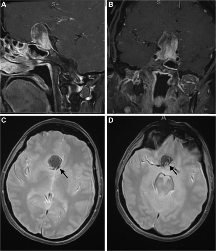
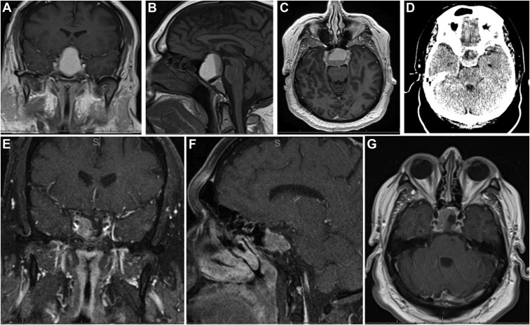
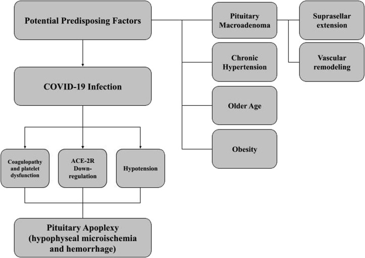
Background: Pituitary apoplexy (PA) is a rare, but life-threatening, condition characterized by pituitary infarction and hemorrhage, most often in the setting of a preexisting adenoma. The risk factors and mechanisms associated with PA are poorly understood. Although neurovascular manifestations of coronavirus disease 2019 (COVID-19) infection have been documented, its association with PA has not yet been determined.
Methods: From a prospectively collected database of patients treated at a tertiary care center for pituitary adenoma, we conducted a retrospective medical record review of PA cases during the COVID-19 pandemic from March 2020 to December 2020. We also conducted a literature review to identify other reported cases.
Results: We identified 3 consecutive cases of PA and concomitant COVID-19 infection. The most common symptoms at presentation were headache and vision changes. The included patients were successfully treated with surgical decompression and medical management of the associated endocrinopathy, ultimately experiencing improvement in their visual symptoms at the latest follow-up examination. COVID-19 infection in the perioperative period was corroborated by polymerase chain reaction test results in all the patients.
Conclusions: With the addition of our series to the literature, 10 cases of PA in the setting of COVID-19 infection have been confirmed. The present series was limited in its ability to draw conclusions about the relationship between these 2 entities. However, COVID-19 infection might represent a risk factor for the development of PA. Further studies are required.
Keywords: COVID-19; Coronavirus; Neurosurgery; Pituitary adenoma; Pituitary apoplexy.
Copyright © 2021 Elsevier Inc. All rights reserved.
Figures

Comment in
-
Letter to the Editor Regarding "Coronavirus Disease 2019 and Pituitary Apoplexy: A Single-Center Case Series and Review of the Literature".World Neurosurg. 2022 Jan;157:252-253. doi: 10.1016/j.wneu.2021.09.023. World Neurosurg. 2022. PMID: 34929775 Free PMC article. No abstract available.
Similar articles
-
Endoscopic Endonasal Surgery for Pituitary Apoplexy: Evidence On a 75-Case Series From a Tertiary Care Center.World Neurosurg. 2017 Oct;106:331-338. doi: 10.1016/j.wneu.2017.06.117. Epub 2017 Jun 30. World Neurosurg. 2017. PMID: 28669873
-
Endoscopic Endonasal Surgery for Treatment of Pituitary Apoplexy: 16 Years of Experience in a Specialized Pituitary Center.World Neurosurg. 2017 Dec;108:137-142. doi: 10.1016/j.wneu.2017.08.131. Epub 2017 Sep 1. World Neurosurg. 2017. PMID: 28867316
-
SARS-CoV-2 Infection, A Risk Factor for Pituitary Apoplexy? A Case Series and Literature Review.Ear Nose Throat J. 2024 Jun;103(1_suppl):153S-161S. doi: 10.1177/01455613231179714. Epub 2023 Jun 8. Ear Nose Throat J. 2024. PMID: 37291861 Free PMC article. Review.
-
Rathke Cleft Cysts with Apoplexy-Like Symptoms: Clinicoradiologic Comparisons with Pituitary Adenomas with Apoplexy.World Neurosurg. 2020 Oct;142:e1-e9. doi: 10.1016/j.wneu.2020.03.086. Epub 2020 Mar 23. World Neurosurg. 2020. PMID: 32217176
-
Pituitary Apoplexy: A Comprehensive Review.Neurol India. 2020 May-Jun;68(Supplement):S72-S78. doi: 10.4103/0028-3886.287669. Neurol India. 2020. PMID: 32611895 Review.
Cited by
-
Newly diagnosed autoimmune Addison's disease in a patient with COVID-19 with autoimmune disseminated encephalomyelitis.BMJ Case Rep. 2022 Dec 5;15(12):e250749. doi: 10.1136/bcr-2022-250749. BMJ Case Rep. 2022. PMID: 36593594 Free PMC article.
-
Case report: Giant pituitary neuroendocrine tumor presented along with acute visual loss due to pituitary apoplexy after receiving COVID-19 vaccination.Front Surg. 2023 Jul 27;10:1220098. doi: 10.3389/fsurg.2023.1220098. eCollection 2023. Front Surg. 2023. PMID: 37576925 Free PMC article.
-
Hypopituitarism and COVID-19.Pituitary. 2024 Dec;27(6):925-934. doi: 10.1007/s11102-024-01463-3. Epub 2024 Nov 19. Pituitary. 2024. PMID: 39560821 Review.
-
Pituitary Apoplexy Secondary to Thrombocytopenia due to Severe Acute Respiratory Syndrome Coronavirus 2 Infection: Report of a Rare Case and Literature Review.J Curr Ophthalmol. 2022 Nov 30;34(3):364-368. doi: 10.4103/joco.joco_321_21. eCollection 2022 Jul-Sep. J Curr Ophthalmol. 2022. PMID: 36644472 Free PMC article.
-
Letter to the Editor Regarding "Coronavirus Disease 2019 and Pituitary Apoplexy: A Single-Center Case Series and Review of the Literature".World Neurosurg. 2022 Jan;157:252-253. doi: 10.1016/j.wneu.2021.09.023. World Neurosurg. 2022. PMID: 34929775 Free PMC article. No abstract available.
References
-
- Johns Hopkins University of Medicine Coronavirus Resource Center. https://coronavirus.jhu.edu/ Available at:
-
- Carod Artal F.J. Complicaciones neurológicas por coronavirus y COVID-19. Rev Neurol. 2020;70:311. - PubMed
Publication types
MeSH terms
Supplementary concepts
LinkOut - more resources
Full Text Sources
Medical
