Current Applications and Selected Technical Details of Dual-Energy X-Ray Absorptiometry
- PMID: 34131097
- PMCID: PMC8216008
- DOI: 10.12659/MSM.930839
Current Applications and Selected Technical Details of Dual-Energy X-Ray Absorptiometry
Abstract
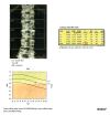
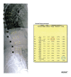
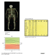
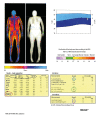
The application of dual-energy X-ray absorptiometry (DXA) examinations in the assessment of bone mineral density (BMD) in the lumbar spine, hip, and forearm is the basic diagnostic method for recognition of osteoporosis. The constant development of DXA technique is due to the aging of societies and the increasing importance of osteoporosis as a public health problem. In order to assess the degree of bone demineralization in patients with hyperparathyroidism, forearm DXA examination is recommended. The vertebral fracture assessment (VFA) of the thoracic and lumbar spine, performed by a highly-skilled technician, is an interesting alternative to the X-ray examination. The DXA total body examination can be useful in the evaluation of fat redistribution among patients after bariatric surgery, in patients infected with HIV and receiving antiretroviral therapy, and in patients with metabolic diseases and suspected to have sarcopenia. The assessment of visceral adipose tissue (VAT) and detection of abdominal aortic calcifications may be useful in the prediction of cardiovascular events. The positive effect of anti-resorptive therapy may affect some parameters of DXA hip structure analysis (HSA). Long-term anti-resorptive therapy, especially with the use of bisphosphonates, may result in changes in the DXA image, which may herald atypical femur fractures (AFF). Reduction of the periprosthetic BMD in the DXA measurements can be used to estimate the likelihood of loosening the prosthesis and periprosthetic fractures. The present review aims to present current applications and selected technical details of DXA.
Conflict of interest statement
None.
Figures


Similar articles
-
Vertebral fracture assessment by dual-energy X-ray absorptiometry along with bone mineral density in the evaluation of postmenopausal osteoporosis.Arch Osteoporos. 2020 Feb 24;15(1):25. doi: 10.1007/s11657-020-0688-9. Arch Osteoporos. 2020. PMID: 32095943
-
Quantitative computed tomography of the lumbar spine, not dual x-ray absorptiometry, is an independent predictor of prevalent vertebral fractures in postmenopausal women with osteopenia receiving long-term glucocorticoid and hormone-replacement therapy.Arthritis Rheum. 2002 May;46(5):1292-7. doi: 10.1002/art.10277. Arthritis Rheum. 2002. PMID: 12115236 Clinical Trial.
-
Bone mineral density at different sites and vertebral fractures in Serbian postmenopausal women.Climacteric. 2017 Feb;20(1):37-43. doi: 10.1080/13697137.2016.1253054. Epub 2016 Nov 18. Climacteric. 2017. PMID: 27860483
-
DXA parameters: beyond bone mineral density.Joint Bone Spine. 2013 May;80(3):265-9. doi: 10.1016/j.jbspin.2012.09.025. Epub 2013 Apr 23. Joint Bone Spine. 2013. PMID: 23622733 Review.
-
Bone densitometry: patients with end-stage renal disease.Health Technol Assess (Rockv). 1996 Mar;(8):1-27. Health Technol Assess (Rockv). 1996. PMID: 8722234 Review.
Cited by
-
Using QCT for the prediction of spontaneous age- and gender-specific thoracolumbar vertebral fractures and accompanying distant vertebral fractures.BMC Musculoskelet Disord. 2024 Oct 19;25(1):828. doi: 10.1186/s12891-024-07961-6. BMC Musculoskelet Disord. 2024. PMID: 39427113 Free PMC article.
-
Quantitative and Qualitative Radiological Assessment of Sarcopenia and Cachexia in Cancer Patients: A Systematic Review.J Pers Med. 2024 Feb 24;14(3):243. doi: 10.3390/jpm14030243. J Pers Med. 2024. PMID: 38540985 Free PMC article. Review.
-
The Significance of Dual-Energy X-ray Absorptiometry (DXA) Examination in Cushing's Syndrome-A Systematic Review.Diagnostics (Basel). 2023 Apr 28;13(9):1576. doi: 10.3390/diagnostics13091576. Diagnostics (Basel). 2023. PMID: 37174967 Free PMC article. Review.
-
Enhancing screening rates for bone health management in prostate cancer patients on androgen deprivation therapy with an automated outpatient system.Sci Rep. 2024 Nov 18;14(1):28460. doi: 10.1038/s41598-024-79888-w. Sci Rep. 2024. PMID: 39558010 Free PMC article.
-
The essential role of dual-energy x-ray absorptiometry in the prediction of subclinical cardiovascular disease.Front Cardiovasc Med. 2024 Aug 29;11:1377299. doi: 10.3389/fcvm.2024.1377299. eCollection 2024. Front Cardiovasc Med. 2024. PMID: 39280034 Free PMC article. Review.
References
-
- Kanis JA, Glüer CC. An update on the diagnosis and assessment of osteoporosis with densitometry. Committee of Scientific Advisors, International Osteoporosis Foundation. Osteoporos Int. 2000;11(3):192–202. - PubMed
-
- Glüer CC. 30 years of DXA technology innovations. Bone. 2017;104:7–12. - PubMed
-
- Storm T, Thamsborg G, Steiniche T, et al. Effect of intermittent cyclical etidronate therapy on bone mass and fracture rate in women with postmenopausal osteoporosis. N Engl J Med. 1990;322:1265–71. - PubMed
-
- Toombs RJ, Ducher G, Shepherd JA, et al. The impact of recent technological advances on the trueness and precision of DXA to assess body composition. Obesity (Silver Spring) 2012;20(1):30–39. - PubMed
Publication types
MeSH terms
LinkOut - more resources
Full Text Sources

