. 2021 Jun 15;12(1):3641.
doi: 10.1038/s41467-021-23911-5.

Generation of patterned kidney organoids that recapitulate the adult kidney collecting duct system from expandable ureteric bud progenitors

Affiliations

Generation of patterned kidney organoids that recapitulate the adult kidney collecting duct system from expandable ureteric bud progenitors

Zipeng Zeng et al. Nat Commun. .

Abstract

Current kidney organoids model development and diseases of the nephron but not the contiguous epithelial network of the kidney's collecting duct (CD) system. Here, we report the generation of an expandable, 3D branching ureteric bud (UB) organoid culture model that can be derived from primary UB progenitors from mouse and human fetal kidneys, or generated de novo from human pluripotent stem cells. In chemically-defined culture conditions, UB organoids generate CD organoids, with differentiated principal and intercalated cells adopting spatial assemblies reflective of the adult kidney's collecting system. Aggregating 3D-cultured nephron progenitor cells with UB organoids in vitro results in a reiterative process of branching morphogenesis and nephron induction, similar to kidney development. Applying an efficient gene editing strategy to remove RET activity, we demonstrate genetically modified UB organoids can model congenital anomalies of kidney and urinary tract. Taken together, these platforms will facilitate an enhanced understanding of development, regeneration and diseases of the mammalian collecting duct system.


Conflict of interest statement

Patent application has been filed on April 27, 2020 by University of Southern California on behalf of inventors Z.Z., B.H., A.P.M., and Z.L. for the mouse and human UB organoid generation, expansion, and CD organoid differentiation systems described in this study. The other authors declare no competing interests.

Figures

Fig. 1
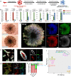
Fig. 1. Expanding mouse UB progenitor cells into 3D branching UB organoids.
a Schematic of mouse UB isolation and screening for UB organoid culture condition. b Representative bright field (BF, left panel) and Wnt11-RFP (right panel) images of UB organoid. Scale bars, 200 µm. c Cumulative growth curve of UB organoid culture starting from 2000 cells. Each time point represents three biological replicates. d Whole-mount immunostaining of UB organoid for various UB markers at Day 10 of culture. The four panels on the right represent the boxed region in the left panel. Scale bars, 100 µm. e Quantification of percentages of UB cells stained positive for different UB markers in Fig. 1d and Supplementary Fig. 2a, b. Each column represents counts from three different fields of view (n = 3). f Principal component analysis (PCA) of RNA-seq data. Different colors and oval circles represent different primary kidney cell populations (NPC, IPC, UB tip, and UB trunk) or UB organoids cultured for 5 (D5), 10 (D10), and 20 days (D20). g Summary of UB organoid derivation from mouse strains with different genetic backgrounds or with different derivation methods. h Bright field (BF) images showing single UB cells, derived from Wnt11-RFP mice, cultured in the UBCM on Days 1 (left panel) and 5 (middle panel), as well as Wnt11-RFP image on Day 5 (right panel). Scale bars, 200 µm. i UB organoid derivation from a single UB cell-derived budding structure isolated from the boxed region in (h) at Days 0 (D0, the day of isolation and re-embedding into Matrigel), 2 (D2), 4 (D4), and 6 (D6). All images in (i) have been scaled to share the same scale bar with the D6 image. Scale bar, 400 µm. All data are presented as mean ± s.d. Source data are provided as a Source Data file.
Fig. 2
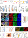
Fig. 2. Generating mature and highly organized CD organoids from UB organoids.
a Schematic of the screening strategy for identifying differentiation culture condition to generate CD organoids from UB organoids. b qRT-PCR analyses of UB (red) and CD (green) organoids for UB progenitor markers Wnt11 and Ret; PC markers Aqp2 and Aqp3; IC markers Foxi1, Atp6v1b1, Slc4a1, and Slc26a4; and Tfcp2l1 that is expressed in both PC and IC. Adult mouse kidney (blue) was used as control. The significance was determined by two-tailed unpaired Student’s t tests. n = 3. c Bright field images showing the morphologic differences between UB and CD organoids. dg Whole-mount immunostaining analyses of CD organoids for PC marker AQP2, and IC markers FOXI1 and ATP6V1B1, showing the distribution of these two cell types within the organoid. f Shows the higher-power images for the boxed region in (e). h Comparison of PC and IC ratios in postnatal Day 0 (P0) mouse CD, adult mouse CD, and CD organoids. Whole-mount immunostaining images (CD organoids), or section staining images (P0 and adult kidneys) stained for AQP2 (PC) and FOXI1 (IC) were quantified for ratios of PC and IC in the CD organoid or the kidney’s collecting duct. Each column represents counts from nine different fields of view (n = 9, three fields were randomly selected from each of the three biologically independent samples). i Principal component analysis (PCA) of RNA-seq data. Different colors and oval circles represent different primary kidney cell populations (NPC, IPC, UB tip, UB trunk, and adult CD cells), or UB organoids cultured for 5 (D5), 10 (D10), and 20 days (D20), or CD organoids. Scale bars: c, d 200 µm, e 100 µm, f 25 µm, g 40 µm. All data are presented as mean ± s.d. Source data are provided as a Source Data file.
Fig. 3
Fig. 3. Generating engineered kidney from expandable NPCs and UBs and gene editing of the UB organoid.
a Schematic of the engineered kidney reconstruction and organotypic culture procedure. b Time course images (bright field and Hoxb7-Venus) showing the branching morphogenesis of the engineered kidney reconstructed at air–liquid interface at Days 2, 4, 5, and 7. Scale bars, 200 µm. c Immunostaining of the engineered kidney (Day 7) constructed from Wnt11-RFP UB organoid and wild-type NPCs for UB/CD marker KRT8, nephron marker PODXL and WT1 (podocytes) and LTL (proximal tubule). Note that both KRT8 and PODXL were stained green. The round structures that co-stain with WT1 are podocytes of the nephron. UB-derived structures do not co-stain with WT1. Scale bar, 100 µm. d Immunostaining of the engineered kidney constructed from Hoxb7-Venus UB organoid and wild-type NPC (Day 10) for GATA3 and CDH1. Scale bars, 50 µm. e Summary of engineered kidney generation experiments. f Schematic of gene overexpression and gene knockout procedures in the UB organoid. OE overexpression, KO knockout. g Fluorescence image of GFP overexpression (GFP OE) in wild-type UB organoid. Scale bar, 200 µm. h Knockout of GFP in Rosa26-Cas9/GFP UB organoid using multiplexed sgRNAs (“GFP KO,” right panels) targeting the coding sequence of GFP. Multiplexed non-targeting sgRNAs were introduced to the organoid as control (“Ctrl KO,” left panel). Note the gene-edited single cells self-organized into typical branching organoid morphology. Scale bars, 400 µm.
Fig. 4
Fig. 4. Generating human UB and CD organoids from primary human UPCs and dual-reporter human pluripotent stem cells.
a Schematic of the purification of primary human UPCs from the nephrogenic zone (illustrated as boxed region) of the human fetal kidney (9–13 weeks of gestational age) and the derivation of human UB organoid. b Immunostaining of the human fetal kidney nephrogenic zone for UB tip marker RET (red), broad UB lineage marker KRT8 (green), and NPC marker SIX2 (cyan). Scale bars, 50 µm. c Time course bright field images showing the growth of human UB organoid derived from primary human UPCs in a typical passage cycle at Days 1 (D1), 5 (D5), and 9 (D9). Scale bar, 200 µm. All images in (c) have been scaled to share the same scale bar with the D9 image. d qRT-PCR analyses of human UB organoid (cultured for 54 days) derived from primary human UPCs for various UB markers as indicated. Human fetal kidney from 11.2-week (11.2 weeks) gestational age was used as control. e Schematic of the stepwise differentiation from WNT11-GFP/PAX2-mCherry dual-reporter hESC line into iUB and iCD organoid. (TeSR mTeSR1 medium, Y Y27632, ME mesendoderm stage medium, UB-I UB Stage I medium, UB-II UB Stage II medium). f qRT-PCR analyses of the FACS purified mCherry+ cells (orange) and the iUB organoid (blue, cultured for 50 days) for various UB markers as indicated. Undifferentiated H1 hESCs (gray) and human fetal kidney (green, 11.2-week gestational age) were used as controls. g Whole-mount immunostaining of the expandable iUB organoid for UB markers SOX9 and CDH1. Scale bars, 50 µm. h Bright field (BF) and PAX2-mCherry (PAX2-mCh) images of expandable iUB organoid (left panels) and mature iCD organoid (right panels). Scale bars, 200 µm. i qRT-PCR analyses of the iUB organoid (orange, cultured for 49 days) and iUB-derived iCD organoid (blue), for UB (WNT11, ETV5), PC (AQP2, AQP4), and IC markers (FOXI1). j Summary of human UB organoid derivation from different sources and their expansion in vitro. ***, this is the culture time we achieved before our lab shutdown due to coronavirus outbreak. The maximum organoid culture time and expansion could be much greater. All data are presented as mean ± s.d. In d, f, i, the significance was determined by two-tailed unpaired Student’s t tests; NS not significant; n = 3. Source data are provided as a Source Data file.
Fig. 5
Fig. 5. Generating human iUB and iCD organoids from human pluripotent stem cells independent of reporters.
a Schematic of the stepwise differentiation from any hPSC line into iUB and iCD organoid without using reporter. (TeSR mTeSR1 medium, CR CloneR, ME (v2) mesendoderm stage medium version 2, UB-I UB Stage I medium, UB-II UB Stage II medium). bh Characterizations of iUB or iCD organoids derived from the WNT11-GFP/PAX2-mCherry hESC line independent of its reporters. b qRT-PCR analyses of the FACS purified KIT+ precursor (orange), KIT+ precursor-derived iUB organoids cultured for 33 (D33, gray), 49 (D49, yellow), and 66 days (D66, light blue) for various UB markers as indicated. Undifferentiated H1 hESCs (dark blue) and human fetal kidney (green, 11.2-week gestational age) were used as controls. ce Whole-mount immunostaining of the expandable iUB organoid for various UB markers as indicated. The four panels on the right represent the boxed region in the left panel. Scale bars, left panel, 100 µm; right four panels, 40 µm. f Quantification of percentages of iUB cells stained positive for different UB markers in Fig. 5c–e. Each column represents counts from three different fields of view (n = 3). g qRT-PCR analyses of the iUB organoid (blue) and iUB-derived iCD organoid (orange), for UB (WNT11, RET), PC (AQP2, AQP3, AQP4), and IC markers (FOXI1). h Whole-mount immunostaining of the iUB-derived iCD organoid for PC marker AQP3 and broad CD marker CDH1. Scale bars, 50 µm. i Flow cytometry analysis of KIT+ precursor cells differentiated from wild-type H1 hESC. j Bright field image of a typical iUB derived from wild-type H1 hESC. Scale bar, 250 µm. k qRT-PCR analyses of the iUB organoids derived from the dual-reporter hESC line (W/P reporter, orange, cultured for 33 days) or wild-type H1 hESC line (H1, gray, cultured for 30 days) for various UB markers. Undifferentiated H1 hESCs (blue) and human fetal kidney (yellow, 11.2-week gestational age) were used as controls. All data are presented as mean ± s.d. In b, g, k, the significance was determined by two-tailed unpaired Student’s t tests; n = 3. Source data are provided as a Source Data file.
Fig. 6
Fig. 6. Modeling kidney development and disease using mouse and human UB organoids.
a Schematic of Ret/RET gene knockout procedures in the mouse or human UB organoid. b Bright field images showing the branching morphogenesis of mouse UB organoids 2 days (Day 2) and 6 days (Day 6) after lentiviral infection. Scale bars, 200 µm. c Whole-mount immunostaining of the control or Ret KO mouse UB organoids for UB markers GATA3 and RET after 6 days of lentiviral infection. Arrow heads indicate the few RET+ cells in the Ret KO organoids. Lower panels represent the boxed region in the upper panels. Scale bars, upper panels, 200 µm; lower panels, 40 µm. d Quantification of percentages of cells stained positive for RET in Fig. 6c. Each column represents counts from three different fields of view (n = 3). e qRT-PCR analyses of the control mouse UB organoids (blue and orange) and Ret KO mouse UB organoids (gray and yellow) for various UB markers 6 days after lentiviral infection. f Bright field images showing the branching morphogenesis of human iUB organoids 3 (Day 3) and 12 days (Day 12) after lentiviral infection. Scale bars, 200 µm. g Whole-mount immunostaining of the control or RET KO human iUB organoids for UB markers PAX2 and RET 12 days after lentiviral infection. Arrow heads indicate the few RET+ cells in the RET KO organoids. Scale bars, 40 µm. See also Supplementary Fig. 7a–f for images of individual fluorescent channels. h Quantification of percentages of human iUB cells stained positive for RET in Fig. 6g. Each column represents counts from three different fields of view (n = 3). i qRT-PCR analyses of the control human iUB organoids (blue and orange) and RET KO iUB organoids (gray and yellow) for various UB markers as indicated 12 days after lentiviral infection. All data are presented as mean ± s.d. In d, e, h, i, the significance was determined by two-tailed unpaired Student’s t tests; n = 3. Source data are provided as a Source Data file.

References

    1. Costantini F, Kopan R. Patterning a complex organ: branching morphogenesis and nephron segmentation in kidney development. Dev. Cell. 2010;18:698–712. doi: 10.1016/j.devcel.2010.04.008. - DOI - PMC - PubMed
    1. Costantini F. Genetic controls and cellular behaviors in branching morphogenesis of the renal collecting system. Wiley Interdiscip. Rev. Dev. Biol. 2012;1:693–713. doi: 10.1002/wdev.52. - DOI - PMC - PubMed
    1. Little, M. H. & McMahon, A. P. Mammalian kidney development: principles, progress, and projections. Cold Spring Harb. Perspect. Biol. 4, 10.1101/cshperspect.a008300 (2012). - PMC - PubMed
    1. McMahon AP. Development of the Mammalian Kidney. Curr. Top. Dev. Biol. 2016;117:31–64. doi: 10.1016/bs.ctdb.2015.10.010. - DOI - PMC - PubMed
    1. Nicolaou N, Renkema KY, Bongers EM, Giles RH, Knoers NV. Genetic, environmental, and epigenetic factors involved in CAKUT. Nat. Rev. Nephrol. 2015;11:720–731. doi: 10.1038/nrneph.2015.140. - DOI - PubMed

Publication types

MeSH terms