. 2021 Aug;22(2):822.
doi: 10.3892/etm.2021.10254. Epub 2021 Jun 2.

Dracorhodin perchlorate enhances wound healing via β-catenin, ERK/p38, and AKT signaling in human HaCaT keratinocytes

Affiliations

Dracorhodin perchlorate enhances wound healing via β-catenin, ERK/p38, and AKT signaling in human HaCaT keratinocytes

Chi-Cheng Lu et al. Exp Ther Med. 2021 Aug.

Abstract

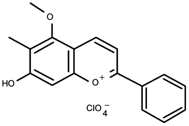
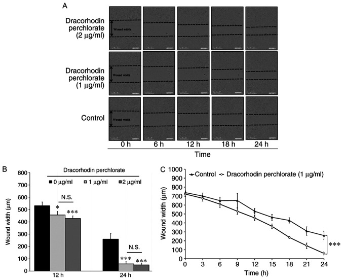
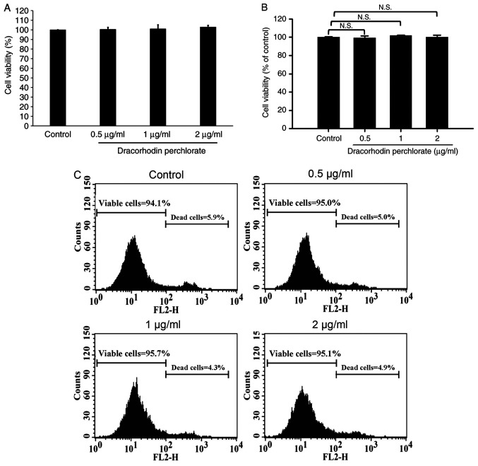
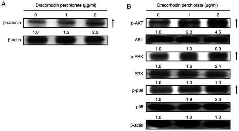
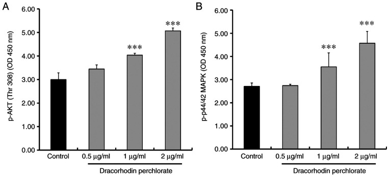
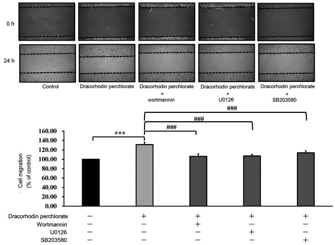
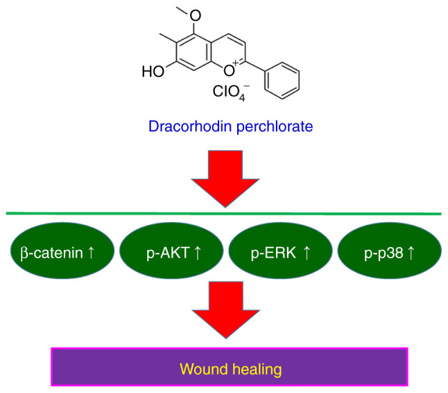
Dracorhodin can be isolated from the exudates of the fruit of Daemonorops draco. Previous studies suggested that dracorhodin perchlorate can promote fibroblast proliferation and enhance angiogenesis during wound healing. In the present study, the potential bioactivity of dracorhodin perchlorate in human HaCaT keratinocytes, were investigated in vitro, with specific focus on HaCaT wound healing. The results of in vitro scratch assay demonstrated the progressive closure of the wound after treatment with dracorhodin perchlorate in a time-dependent manner. An MTT assay and propidium iodide exclusion detected using flow cytometry were used to detect cell viability of HaCaT cells. Potential signaling pathways underlying the effects mediated by dracorhodin perchlorate in HaCaT cells were clarified by western blot analysis and kinase activity assays. Dracorhodin perchlorate significantly increased the protein expression levels of β-catenin and activation of AKT, ERK and p38 in HaCaT cells. In addition, dracorhodin perchlorate did not induce HaCaT cell proliferation but promoted cell migration. Other mechanisms may yet be involved in the dracorhodin perchlorate-induced wound healing process of human keratinocytes. In summary, dracorhodin perchlorate may serve to be a potential molecularly-targeted phytochemical that can improve skin wound healing.

Keywords: ERK/p38/AKT signaling; HaCaT keratinocytes; dracorhodin perchlorate. β-catenin; wound healing.


Conflict of interest statement

The authors declare that they have no competing interests.

Figures

Figure 1
Figure 1
Chemical structure of dracorhodin perchlorate.
Figure 2
Figure 2
Determination of cell migration in HaCaT keratinocytes. (A) Effects of dracorhodin perchlorate on wound healing of HaCaT keratinocytes. Cells (1x10⁴ cells/well) in 96-well plates were scratched and incubated with or without 1 and 2 µg/ml dracorhodin perchlorate for indicated time. (B) Cells (1x10⁴ cells/well) in a 96-well plate were scratched and treated with or without 1 and 2 µg/ml dracorhodin perchlorate for 12 and 24 h. The wound width of HaCaT keratinocytes was determined using the IncuCyte ZOOM System instrument and then quantified. (C) Time courses of the relative wound widths of HaCaT keratinocytes after wound generation. Data are presented as the mean ± standard deviation, n=3. Tukey's post hoc test after ANOVA. *P<0.05 and ***P<0.001 vs. 0 µg/ml untreated control. N.S., not significant.
Figure 3
Figure 3
Effects of dracorhodin perchlorate on cell viability in HaCaT keratinocytes. Cells (2.5x10⁵ cells/well) in 24-well plates were incubated with 0, 0.5, 1 and 2 µg/ml dracorhodin perchlorate for 24 h. The cell viability was determined by (A) MTT assay, (B) propidium iodide exclusion method, and (C) a representative histogram of the quantified flow cytometry plots. Data are presented as the mean ± standard deviation, n=3. Dunnett's post hoc test after ANOVA. N.S., not significant.
Figure 4
Figure 4
Effects of dracorhodin perchlorate on the protein expression levels of β-catenin, AKT, ERK and p38 proteins in HaCaT keratinocytes. Cells were incubated with 0, 1 and 2 µg/ml dracorhodin perchlorate for 12 h. Whole cell lysates were prepared before the levels of cell migration-related proteins were analyzed by western blot analysis. The protein levels of (A) β-catenin, (B) p-AKT, AKT, p-ERK, ERK, p-p38 and p38 were detected. All blots were normalized to β-actin to ensure equal loading.
Figure 5
Figure 5
Effects of dracorhodin perchlorate on the AKT and ERK kinase activities in HaCaT keratinocytes. Cells were incubated with 0, 0.5, 1 and 2 µg/ml dracorhodin perchlorate for 12 h. Whole cell lysates were then prepared and the (A) AKT and (B) ERK kinase activities were analyzed by sandwich p-AKT and p-ERK ELISA. Data are presented as the mean ± standard deviation, n=3. Dunnett's post hoc test after ANOVA. ***P<0.001 vs. Control. OD, optical density.
Figure 6
Figure 6
Effect of protein kinase inhibitors on the cell migration of dracorhodin perchlorate-treated keratinocytes. Cells were incubated with or without dracorhodin perchlorate (1 µg/ml) and three protein kinase inhibitors for 24 h. Wortmannin (10 µM), U0126 (10 µM) and SB203580 (10 µM) are AKT, ERK, and p38 inhibitors, respectively. Data are presented as the mean ± standard deviation, n=3. Tukey's post hoc test after ANOVA. ***P<0.001 and ###P<0.001.
Figure 7
Figure 7
Summary of the possible mechanism underlying the wound healing of dracorhodin perchlorate in HaCaT keratinocytes.

References

    1. Pona A, Cline A, Kolli SS, Taylor SL, Feldman SR. Review of future insights of Dragon's Blood in dermatology. Dermatol Ther. 2019;32(e12786) doi: 10.1111/dth.12786. - DOI - PubMed
    1. Sousa MM, Melo MJ, Parola AJ, Seixas de Melo JS, Catarino F, Pina F, Cook FE, Simmonds MS, Lopes JA. Flavylium chromophores as species markers for dragon's blood resins from Dracaena and Daemonorops trees. J Chromatogr A. 2008;1209:153–161. doi: 10.1016/j.chroma.2008.09.007. - DOI - PubMed
    1. Edwards HG, de Oliveira LF, Prendergast HD. Raman spectroscopic analysis of dragon's blood resins-basis for distinguishing between Dracaena (Convallariaceae), Daemonorops (Palmae) and Croton (Euphorbiaceae) Analyst. 2004;129:134–138. doi: 10.1039/b311072a. - DOI - PubMed
    1. Wu C, Cai XQ, Chang Y, Chen CH, Ho TJ, Lai SC, Chen HP. Rapid identification of dragon blood samples from Daemonorops draco, Dracaena cinnabari and Dracaena cochinchinensis by MALDI-TOF mass spectrometry. Phytochem Anal. 2019;30:720–726. doi: 10.1002/pca.2852. - DOI - PubMed
    1. Al-Okaishi A. Exploring the historical distribution of Dracaena cinnabari using ethnobotanical knowledge on Socotra Island, Yemen. J Ethnobiol Ethnomed. 2021;17(22) doi: 10.1186/s13002-021-00452-1. - DOI - PMC - PubMed