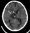
Chordoid glioma in the thalamus of a child: Rare location and atypical imaging findings
- PMID: 34131491
- PMCID: PMC8171145
- DOI: 10.1259/bjrcr.20200108
Chordoid glioma in the thalamus of a child: Rare location and atypical imaging findings
Abstract
Chordoid glioma is a rare intracranial tumour, which usually occurs in middle-aged female patients, mainly in the third ventricle, hypothalamus and suprasellar region. It can reportedly occur in the temporal-parietal lobe, occipital horn of the lateral ventricle and left thalamus. Here, we report a case of chordoid glioma in the thalamic region of a female child, which is different from the previously reported chordoid glioma in the left thalamus. Given its atypical location and imaging findings, it is often misdiagnosed as low-grade glioma before operation. Through the study of this case, we recognized the atypical imaging manifestations of chordoid glioma in a rare location.
© 2020 The Authors. Published by the British Institute of Radiology.
Conflict of interest statement
Conflict of interest: The authors report no conflict of interest concerning the materials or methods used in this study, or related to the findings specified in this paper.
Figures


Similar articles
-
Chordoid glioma: report of two rare examples with unusual features.Acta Neurochir (Wien). 2008 Mar;150(3):295-300; discussion 300. doi: 10.1007/s00701-008-1420-x. Epub 2008 Feb 4. Acta Neurochir (Wien). 2008. PMID: 18246456
-
Chordoid glioma : a case report of unusual location and neuroradiological characteristics.J Korean Neurosurg Soc. 2010 Jul;48(1):62-5. doi: 10.3340/jkns.2010.48.1.62. Epub 2010 Jul 31. J Korean Neurosurg Soc. 2010. PMID: 20717514 Free PMC article.
-
Chordoid glioma of the third ventricle: a patient presenting with SIADH and a review of this rare tumor.Pituitary. 2016 Aug;19(4):356-61. doi: 10.1007/s11102-016-0711-8. Pituitary. 2016. PMID: 26879322 Review.
-
Chordoid glioma: a neoplasm unique to the hypothalamus and anterior third ventricle.AJNR Am J Neuroradiol. 2001 Mar;22(3):464-9. AJNR Am J Neuroradiol. 2001. PMID: 11237967 Free PMC article.
-
Chordoid glioma of the third ventricle: a report of two new cases, with further evidence supporting an ependymal differentiation, and review of the literature.Am J Surg Pathol. 2002 Oct;26(10):1330-42. doi: 10.1097/00000478-200210000-00010. Am J Surg Pathol. 2002. PMID: 12360048 Review.
References
Publication types
LinkOut - more resources
Full Text Sources

