

Incidental cardiac uptake in bone scintigraphy: increased importance and association with cardiac amyloidosis
- PMID: 34131495
- PMCID: PMC8171131
- DOI: 10.1259/bjrcr.20200161
Incidental cardiac uptake in bone scintigraphy: increased importance and association with cardiac amyloidosis
Abstract
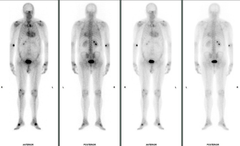
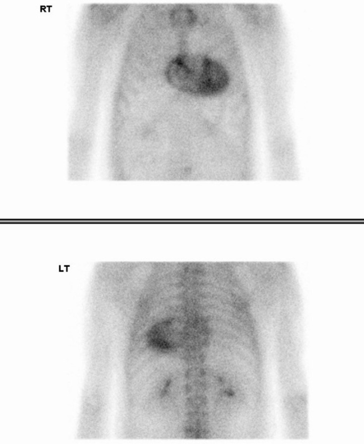
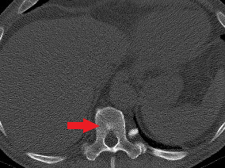
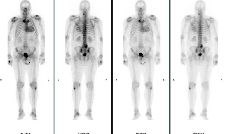
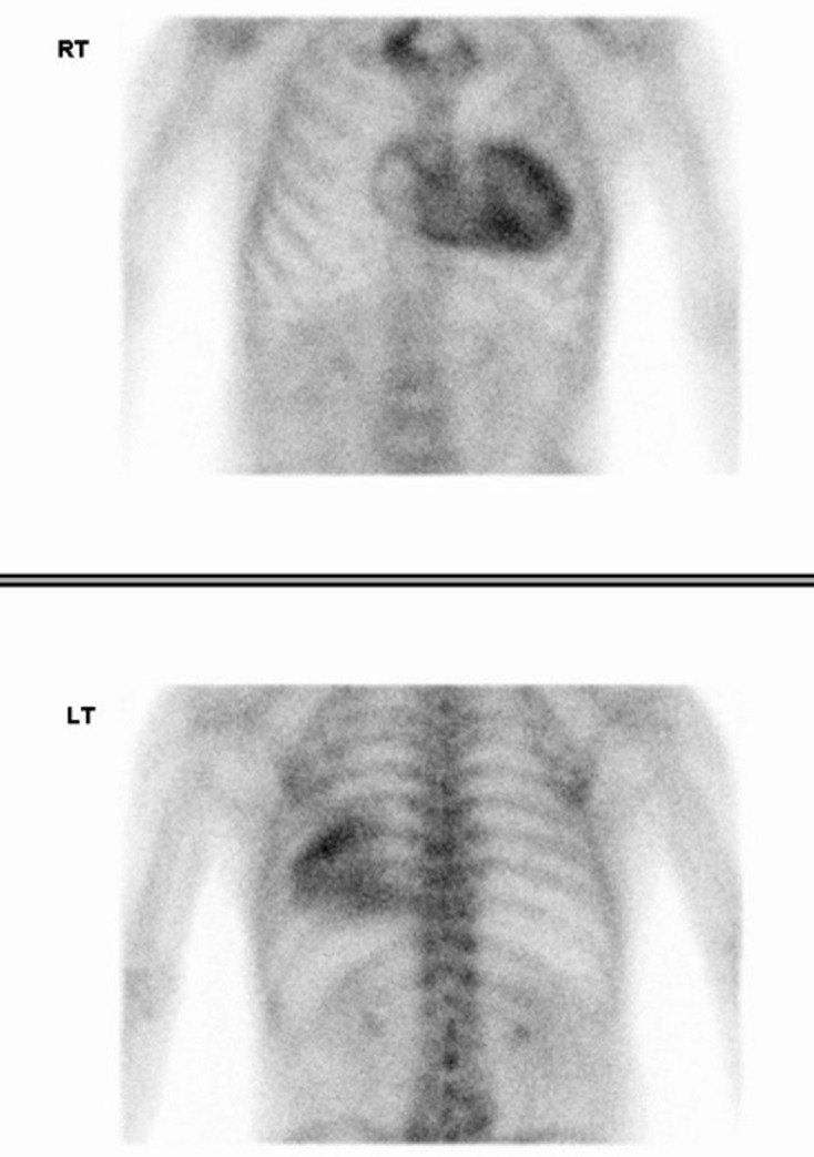
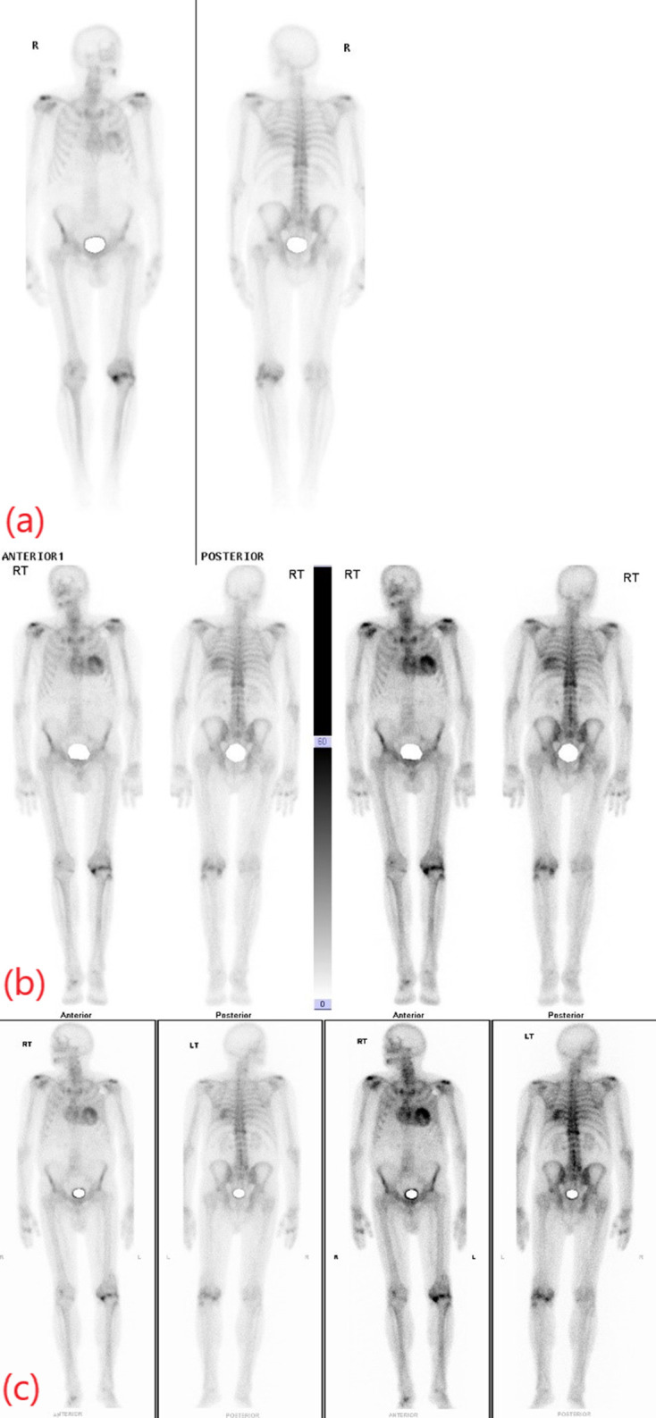
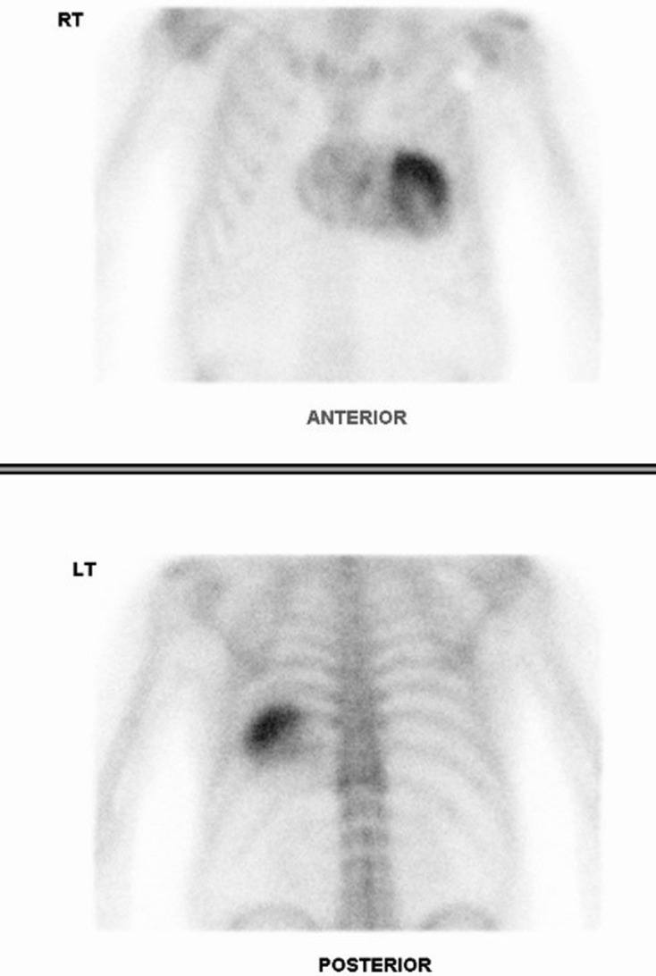
Extraosseous radiotracer uptake during bone scintigraphy must be carefully assessed and it offers the potential to detect previously undiagnosed disease processes. A range of neoplastic, metabolic, traumatic, ischaemic and inflammatory disorders can cause soft tissue accumulation of bone avid radiopharmaceuticals. Accordingly, cardiac uptake in bone scintigraphy has a broad differential diagnosis and is commonly attributed to ischaemia/infarction related to coronary artery disease. However, there has been renewed focus on incidental cardiac uptake in recent years in light of significant developments in the diagnosis and management of cardiac amyloidosis.
© 2021 The Authors. Published by the British Institute of Radiology.
Figures
Similar articles
-
Noninvasive etiologic diagnosis of cardiac amyloidosis using 99mTc-3,3-diphosphono-1,2-propanodicarboxylic acid scintigraphy.J Am Coll Cardiol. 2005 Sep 20;46(6):1076-84. doi: 10.1016/j.jacc.2005.05.073. J Am Coll Cardiol. 2005. PMID: 16168294
-
Bone scintigraphy with (99m)technetium-hydroxymethylene diphosphonate allows early diagnosis of cardiac involvement in patients with transthyretin-derived systemic amyloidosis.Amyloid. 2014 Mar;21(1):35-44. doi: 10.3109/13506129.2013.871250. Epub 2014 Jan 23. Amyloid. 2014. PMID: 24455993
-
Utility and limitations of 3,3-diphosphono-1,2-propanodicarboxylic acid scintigraphy in systemic amyloidosis.Eur Heart J Cardiovasc Imaging. 2014 Nov;15(11):1289-98. doi: 10.1093/ehjci/jeu107. Epub 2014 Jun 16. Eur Heart J Cardiovasc Imaging. 2014. PMID: 24939945
-
Clinical Phenotyping of Transthyretin Cardiac Amyloidosis with Bone-Seeking Radiotracers in Heart Failure with Preserved Ejection Fraction.Curr Cardiol Rep. 2018 Mar 8;20(4):23. doi: 10.1007/s11886-018-0970-2. Curr Cardiol Rep. 2018. PMID: 29520480 Review.
-
Targeted Nuclear Imaging Probes for Cardiac Amyloidosis.Curr Cardiol Rep. 2017 Jul;19(7):59. doi: 10.1007/s11886-017-0868-4. Curr Cardiol Rep. 2017. PMID: 28508350 Review.
Cited by
-
World Heart Federation Consensus on Transthyretin Amyloidosis Cardiomyopathy (ATTR-CM).Glob Heart. 2023 Oct 26;18(1):59. doi: 10.5334/gh.1262. eCollection 2023. Glob Heart. 2023. PMID: 37901600 Free PMC article. Review.
-
Unexplained Cardiac Uptake on 99mTc-MDP Bone Scan in a Patient with Prostate Cancer.Asia Ocean J Nucl Med Biol. 2025;13(2):198-202. doi: 10.22038/aojnmb.2025.85251.1609. Asia Ocean J Nucl Med Biol. 2025. PMID: 40585290 Free PMC article.
-
The importance of pathways to facilitate early diagnosis and treatment of patients with cardiac amyloidosis.Ther Adv Cardiovasc Dis. 2023 Jan-Dec;17:17539447231216318. doi: 10.1177/17539447231216318. Ther Adv Cardiovasc Dis. 2023. PMID: 38099406 Free PMC article. Review.
References
Publication types
LinkOut - more resources
Full Text Sources
