Case Reports
. 2021 Jan 6;7(3):20200183.
doi: 10.1259/bjrcr.20200183. eCollection 2021 May 1.

A rare case of paradoxical pulmonary embolism in spontaneous aortocaval fistula

Affiliations
Case Reports

A rare case of paradoxical pulmonary embolism in spontaneous aortocaval fistula

Valentina Vespro et al. BJR Case Rep. .

Abstract

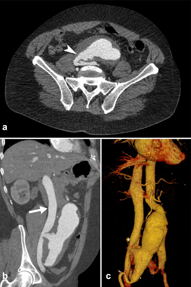
Aortocaval fistula (ACF) is a rare complication of abdominal aortic aneurysm (AAA), occurring in less than 1% of all AAAs. Paradoxical embolism can rarely be associated with ACF, pulmonary embolism may originate from dislodgment of thrombotic material from the AAA in the inferior vena cava (IVC) through the ACF. We report a case of a patient admitted to the emergency department with abdominal pain and shortness of breath who immediately underwent thoraco-abdominal CT. Imaging allowed a prompt pre-operative diagnosis of an ACF between an AAA and the IVC, also identifying CT signs of right heart overload and the presence of a paradoxical pulmonary embolism.


Figures

Figure 1.
Figure 1.
Contrast-enhanced CT scan during arterial-phase showing the presence of a fistula between the aortic aneurysm and the IVC. (a) Axial images well depict the communication between the two vessels (arrowhead). (b, c) MPR and VRT reformatted images show the early full enhancement of the dilated IVC (arrow) during the arterial phase
Figure 2.
Figure 2.
Contrast-enhanced CT scan during arterial-phase. (a) Lower chest CT image demonstrates CT signs of right heart overload: enlargement of right atrium and ventricle, cardiac axis clockwise rotation (curved arrow) and straightened interventricular septum (arrowhead). (b, c, d) Axial and MPR reformatted images showing bilateral segmental and subsegmental pulmonary embolism (arrows)
Figure 3.
Figure 3.
(a) IVC suture at the level of aortocaval fistula. (b) Aorto-bisiliac reconstruction with Dacron ® prosthesis

References

    1. De Rango P, Parlani G, Cieri E, Verzini F, Isernia G, Silvestri V, et al. . Paradoxical pulmonary embolism with spontaneous aortocaval fistula. Ann Vasc Surg 2012; 26: 739–46. doi: 10.1016/j.avsg.2011.06.011 - DOI - PubMed
    1. Tsolakis JA, Papadoulas S, Kakkos SK, Skroubis G, Siablis D, Androulakis JA. Aortocaval fistula in ruptured aneurysms. Eur J Vasc Endovasc Surg 1999; 17: 390–3. doi: 10.1053/ejvs.1998.0777 - DOI - PubMed
    1. Lorenzati B, Perotto M, Bottone S, Tenconi G, Gazzina G, Cataldi W. Aortocaval fistula. Intern Emerg Med 2014; 9: 895–6. doi: 10.1007/s11739-014-1076-5 - DOI - PubMed
    1. Thet Y, Ranjit A, Ravi R, Khand A. High output cardiac failure and paradoxical pulmonary emboli secondary to aortocaval fistula. Postgrad Med J 2012; 88: 613–4. doi: 10.1136/postgradmedj-2012-130840 - DOI - PubMed
    1. Laporte F, Olivier A, Groben L, Admant P, Aliot E. Aortocaval fistula: an uncommon cause of paradoxical embolism. J Cardiovasc Med 2012; 13: 68–71. doi: 10.2459/JCM.0b013e32834039d7 - DOI - PubMed

Publication types