

Infantile mesenchymal hamartoma of the liver with elevated alpha fetoprotein
- PMID: 34131505
- PMCID: PMC8171140
- DOI: 10.1259/bjrcr.20200196
Infantile mesenchymal hamartoma of the liver with elevated alpha fetoprotein
Abstract
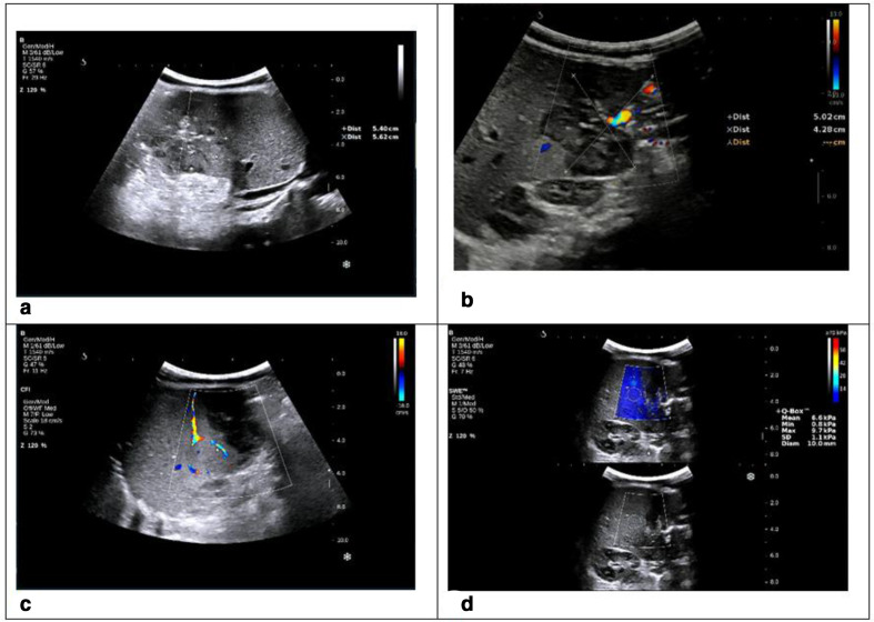
Mesenchymal hamartoma of the liver (MHL) is a benign tumour that most commonly occurs in children. In most cases of MHL, the α fetoprotein (AFP) level is within the normal limits, only in a few cases, increased AFP has been described which usually causes misdiagnosis of hepatoblastoma. We report a case of a 3-month-old paediatric patient who was incidentally detected with a very high level of AFP, at 6388.4 ng ml-1. Ultrasound revealed a right liver tumour, segment VI, measuring at 56 × 53 mm. According to images of ultrasound and MRI, the diagnosis was mesenchymal hepatic sarcoma. The paediatric patient had surgery to remove the entire liver segment containing the tumour. Micropathological examination showed that the tumour was a MHL. The serum AFP level fell rapidly to near normal following the surgery. The MHL benign liver tumour with an atypical presentation caused a very high AFP level. This was a rare clinical case, and it was difficult to diagnose.
© 2021 The Authors. Published by the British Institute of Radiology.
Figures


Similar articles
-
Mesenchymal hamartoma of the liver mimicking hepatoblastoma.J Pediatr Hematol Oncol. 2008 Jun;30(6):458-60. doi: 10.1097/MPH.0b013e318169171b. J Pediatr Hematol Oncol. 2008. PMID: 18525464
-
Fetal and neonatal hepatic tumors.J Pediatr Surg. 2007 Nov;42(11):1797-803. doi: 10.1016/j.jpedsurg.2007.07.047. J Pediatr Surg. 2007. PMID: 18022426 Review.
-
Mesenchymal hamartoma mimicking hepatoblastoma.Int J Organ Transplant Med. 2014;5(2):78-80. Int J Organ Transplant Med. 2014. PMID: 25013683 Free PMC article.
-
Mesenchymal hamartoma of the liver associated with features of Beckwith-Wiedemann syndrome and high serum alpha-fetoprotein levels.Pediatr Dev Pathol. 2007 May-Jun;10(3):233-8. doi: 10.2350/06-07-0128.1. Pediatr Dev Pathol. 2007. PMID: 17535089
-
Distinctive case. Adult mesenchymal hamartoma of the liver: report of a case with light microscopic, FNA cytology, immunohistochemistry, and ultrastructural studies and review of the literature.Mod Pathol. 1991 May;4(3):392-5. Mod Pathol. 1991. PMID: 2068067 Review.
Cited by
-
Mesenchymal hepatic hamartoma: A rare case of severe respiratory distress in a neonate.Clin Case Rep. 2024 Mar 13;12(3):e8562. doi: 10.1002/ccr3.8562. eCollection 2024 Mar. Clin Case Rep. 2024. PMID: 38487637 Free PMC article.
References
-
- Atas E, Demirkaya M, et al. . Mesenchymal hamartoma of the liver mimicking hydatid cyst. Pediatr Therapeut 2012; 2. doi: 10.4172/2161-0665.1000120 - DOI
-
- Gaxa L, Hlatshwayo B. Mesenchymal hepatic hamartoma associated with elevated alpha fetoprotein mimicking a hepatoblastoma: a rare case and a literature review. Case Reports International 2017; 6: 9–12. doi: 10.5348/crint-2017-34-CR-3 - DOI
-
- Chung EM, Cube R, Lewis RB, Conran RM, et al. . From the archives of the AFIP: pediatric liver masses: radiologic-pathologic correlation part 1. benign tumors. Radiographics 2009; 30: 801–26. - PubMed
Publication types
LinkOut - more resources
Full Text Sources
